Review
. 2021 Feb 11;13(2):185-202.
doi: 10.1007/s12551-021-00787-9. eCollection 2021 Apr.

Characterization of immune cell migration using microfabrication

Affiliations
Review

Characterization of immune cell migration using microfabrication

Doriane Vesperini et al. Biophys Rev. .

Abstract

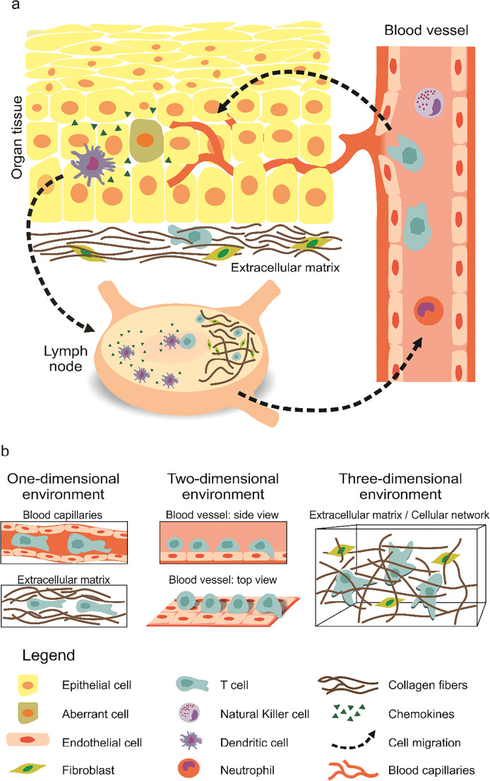
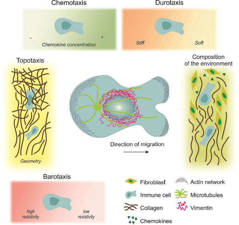
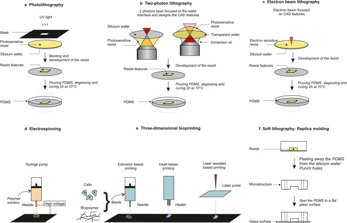
The immune system provides our defense against pathogens and aberrant cells, including tumorigenic and infected cells. Motility is one of the fundamental characteristics that enable immune cells to find invading pathogens, control tissue damage, and eliminate primary developing tumors, even in the absence of external treatments. These processes are termed "immune surveillance." Migration disorders of immune cells are related to autoimmune diseases, chronic inflammation, and tumor evasion. It is therefore essential to characterize immune cell motility in different physiologically and pathologically relevant scenarios to understand the regulatory mechanisms of functionality of immune responses. This review is focused on immune cell migration, to define the underlying mechanisms and the corresponding investigative approaches. We highlight the challenges that immune cells encounter in vivo, and the microfabrication methods to mimic particular aspects of their microenvironment. We discuss the advantages and disadvantages of the proposed tools, and provide information on how to access them. Furthermore, we summarize the directional cues that regulate individual immune cell migration, and discuss the behavior of immune cells in a complex environment composed of multiple directional cues.

Keywords: Amoeboid migration; Immune cells; Microfabrication; Target search.


Conflict of interest statement

Conflict of interestThe authors declare that they have no conflict of interest.

Figures

Fig. 1
Fig. 1
Immune cell migration in vivo and the diverse scenarios encountered. a) Overview of immune cell migration in vivo. From blood vessels, immune cells transmigrate into and then patrol peripheral tissues/organs to clear invaders and/or collect antigens. Then, immune cells enter lymph vessels and migrate toward the lymph nodes. Peripheral dendritic cells (DCs) are responsible to collect antigens from aberrant (infected or malignant) cells in peripheral tissues (skin is shown here). Upon recognition of an invader or aberrant cells, an immediate immune response is initiated locally. Then, professional antigen-presenting cells (APCs) go to the lymph nodes, where the adaptive immune cells (B and T cells) are activated. Activation is then followed by proliferation. Effector cells enter the blood circulation and transmigrate into the respective inflammation sites. b) Schematic of 1D, 2D and 3D scenarios encountered by immune cells during migration. T cells are drawn here in the illustration as an example of immune cells. 1D is found in blood/lymph capillaries and in the cavities/channels in ECM. The blood vessel walls, which immune cells are rolling on, correspond to a 2D scenario. In general, while patrolling the tissues, immune cells face a 3D environment with ECM as a main component
Fig. 2
Fig. 2
Migratory challenges and guidance cues encountered by immune cells during circulation. The extracellular regulation of cell migration includes: chemokines and stiffness gradients; the extracellular matrix (ECM) mechanics (including loose or highly cross-linked zones) and its topolography (pores, or obstacles); the molecular composition of the matrix surrounding the cells (collagen, fibroblasts, chemokines) as well as pressure gradients. Center: Schematic representation of a polarized immune cell migrating directionally in an amoeboid migration mode. Amoeboid migration is characterized by a round cell morphology, low adhesive contacts and cell body deformation driven by actin protrusions. The microtubule organizing center is generally located at the back of the nucleus. The integrity of the nucleus is protected by a nuclear cage formed by intermediate filament proteins, such as vimentin
Fig. 3
Fig. 3
Techniques used to study in vitro cell migration. (a) Photolithography: the photosensitive resist is exposed to UV light through a mask where the features are designed. (b) Two-photon lithography: mask-free technique where the features are directly printed in the resist from the resist–wafer interface with a piezo motion in the three space dimensions (X, Y, Z). The resist is up or down depending on the wafer transparency (image inspired by (Bückmann et al. 2012)). (c) Electron beam lithography: mask-free technique where the features are directly printed by reticulation of the resist in contact with the electron beam. (d) Electrospinning: mask free technique where a polymer solution is extruded from a needle around which a high voltage electric field is formed, and deposited on a surface that can be rotating. The 2D membrane fabricated using this technique can then have random to aligned nanofibrous structures depending on the needle translation. (e) Three-dimensional bioprinting: mask-free technique where a bio-ink consisting of cells and biopolymers is directly deposited on a surface by extrusion-, inkjet- or laser-assisted-based printing. (f) Soft lithography: this step follows the production of the wafer with one of the techniques presented in (a, b, and c); polydimethylsiloxane (PDMS) is peeled away from the wafer, punched, cut, cleaned and bound to a glass surface. CAD, computer-aided design

References

    1. Abdul Hamid AI, Nakusi L, Givskov M, Chang YT, Marques C, Gueirard P. A mouse ear skin model to study the dynamics of innate immune responses against Staphylococcus aureus biofilms. BMC Microbiol. 2020;20:22. doi: 10.1186/s12866-019-1635-z. - DOI - PMC - PubMed
    1. Aizel K, Clark AG, Simon A, Geraldo S, Funfak A, Vargas P, Bibette J, Vignjevic DM, Bremond N. A tuneable microfluidic system for long duration chemotaxis experiments in a 3D collagen matrix. Lab Chip. 2017;17:3851–3861. doi: 10.1039/C7LC00649G. - DOI - PubMed
    1. Altissimo M. E-beam lithography for micro−/nanofabrication. Biomicrofluidics. 2010;4:026503. doi: 10.1063/1.3437589. - DOI - PMC - PubMed
    1. Ambravaneswaran V, Wong IY, Aranyosi AJ, Toner M, Irimia D. Directional decisions during neutrophil chemotaxis inside bifurcating channels. Integ Biol. 2010;2:639–647. doi: 10.1039/c0ib00011f. - DOI - PMC - PubMed
    1. Bajénoff M, Glaichenhaus N, Germain RN. Fibroblastic reticular cells guide T lymphocyte entry into and migration within the splenic T cell zone. J Immunol. 2008;181:3947–3954. doi: 10.4049/jimmunol.181.6.3947. - DOI - PMC - PubMed

LinkOut - more resources