. 2022 Feb;45(1):771-781.
doi: 10.1007/s10143-021-01570-w. Epub 2021 Jul 22.

Endoscopic third ventriculostomy before surgery of third ventricle and posterior fossa tumours decreases the risk of secondary hydrocephalus and early postoperative complications

Affiliations

Endoscopic third ventriculostomy before surgery of third ventricle and posterior fossa tumours decreases the risk of secondary hydrocephalus and early postoperative complications

Pawel Tabakow et al. Neurosurg Rev. 2022 Feb.

Abstract

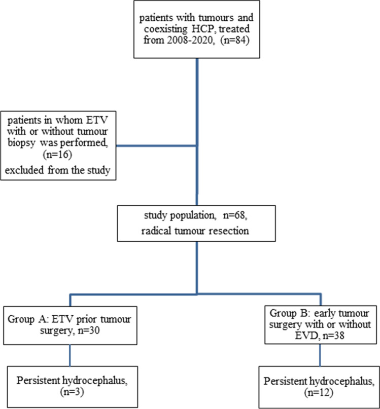
Endoscopic third ventriculostomy (ETV) is an effective treatment for obstructive hydrocephalus (HCP) at the level of third or fourth ventricle. To date, there is no consensus regarding its role as intervention preceding the operation of tumour removal. The aim of this prospective open-label controlled study is to assess if ETV prevents secondary HCP after tumour removal and if ETV influences the early results of tumour surgery. The study was performed on 68 patients operated for tumours of the third ventricle and posterior fossa. In 30 patients, ETV was performed several days before tumour removal, while in 38 patients, the tumour was removed during a one-stage procedure without ETV. Patients who did not receive ETV before the tumour removal procedure had a higher probability of developing postoperative HCP (n = 12, p = 0.03). They also demonstrated a substantially higher rate of early postoperative complications (n = 20, p = 0.002) and a lower Karnofsky score (p = 0.004) than patients in whom ETV was performed before tumour removal. The performance of external ventricular drainage in the non-ETV group did not prevent secondary HCP (p = 0.68). Postoperative cerebellar swelling (p = 0.01), haematoma (p = 0.04), cerebrospinal fluid leak (p = 0.04) and neuroinfection (p = 0.04) were the main risk factors of persistent HCP. Performance of ETV before tumour removal is not only beneficial for control of acute HCP but also prevents the occurrence of secondary postoperative HCP and may also minimize early postoperative complications.

Keywords: Endoscopic third ventriculostomy; Secondary postoperative hydrocephalus; Tumour removal.


Conflict of interest statement

The authors declare no competing interests.

Figures

Fig. 1
Fig. 1
Flow chart describing the study population and the results concerning the primary outcome measure
Fig. 2
Fig. 2
MRI scans performed in a 26-year-old woman with a 4th ventricle subependymoma. (A), (B) and (C) show axial fiesta-3D and contrast-enhanced T1-weighted coronal and sagittal images. Orange arrows show the tumour obliterating the foramina of the 4th ventricle with a subsequent obstructive four-ventricle HCP. (C), (D) and (F) demonstrate the FLAIR axial and T2-weighted coronal and sagittal MRI images after 10 years following ETV and tumour resection. Note that there is no tumour visible and no evidence of HCP (a marked decrease in Evans Index). The blue arrow in picture (F) shows the flow void phenomenon across the area of ventriculostomy, while the green arrow points to areas of possible postoperative adhesions (in black) at the level of the foramen of Magendie
Fig. 3
Fig. 3
A case of a 7-year-old girl operated for pilocytic astrocytoma of the posterior fossa. (A) Sagittal T2-weighted scan shows a large tumour obliterating the 4th ventricle. (B) FLAIR axial scan demonstrates active HCP. (C), (D) Fiesta 3D sagittal and axial scans show the tumour compressing the brain stem and cerebellum, restricting the CSF flow. (E) Postoperative CT scan performed on day 6 after tumour resection. HCP is still present, although the patient had undergone EVD (orange arrow). For this reason, the patient underwent ETV. (F) CT scan from day 6 shows no evidence of tumour in the posterior fossa. However, there is still cerebellar oedema and some CSF collection in the epidural space (orange arrow). (G), (H), (I) MRI scans performed a year after tumour removal. (G) A large collection of CSF through a dural fistula is shown on the axial Fiesta 3D image (orange arrow). (H) Sagittal T2-weighted image shows the pseudomeningocoele (blue arrow), but, at the same time, good flow of CSF through the area of ventriculostomy (orange arrow) and lack of HCP. A pineal cyst is also present. (I) Axial FLAIR scan shows normal ventricle size after ETV performance
Fig. 4
Fig. 4
Comparison of outcomes in a 21-year-old man from group B (AF) and a 51-year-old woman from group A (GN) In both cases, an ependymoma of the 4th ventricle was radically resected, and in both cases, postoperative haematoma was noted. While the course of treatment of patient A was uneventful, the patient from group B continued to deteriorate and died a month after the tumour surgery. (A) Sagittal T2-weighted MRI scans show a large tumour obliterating the posterior part of the 4th ventricle and the craniovertebral junction. (B) CT images performed on day 1 after tumour resection demonstrate a haematoma obliterating the 4th ventricle, the Sylvian aqueduct and partially the 3rd ventricle. For this reason, the EVD was kept longer, but as the patient did not improve and additionally presented CSF leak, on a postoperative day 7, the haematoma was removed, and duraplasty was performed. (CF) CT and MRI scans from day 28 to 29, respectively. In the meantime, the patient underwent 2 additional procedures of EVD replacement and was treated for bacterial meningitis and ventriculitis. (C) While no haematoma is visible intraventricularly, there is a marked cerebellar oedema/ischaemia. CSF fistula is still visible (blue arrow). The ventricles are still dilated despite the presence of a ventricular drain (orange arrow). (D) Axial T1-weighted contrast-enhanced images show HCP and ventricle enhancement due to ventriculitis. (E) FLAIR MRI axial scan demonstrates active HCP (orange arrow) and multiple posthaemorrhagic and postinfectious intraventricular fibrous adhesions (blue arrow). (F) Sagittal T2-weighted MRI scans show further adhesions at the level of the 4th ventricle (orange arrows) and subcutaneous CSF collection, secondary to the active HCP (blue arrow). After removal of EVD, the patient finally underwent endoscopic removal of the intraventricular blood remnants. Some adhesions were removed, including the ones in the aqueduct, and ETV was performed. Unfortunately, the patient died several days later due to neuroinfection. (G) T2-weighted sagittal MRI scan of a patient from group A shows an almost identical 4th ventricle ependymoma. (H) Axial T2-weighted scan demonstrates HCP. (I) CT scan performed after ETV shows some narrowing of the ventricles. The patient underwent tumour resection 9 days after ETV. (J) CT images on day 1 after complete tumour resection show some intraventricular blood. (KL) Axial CT scans (day 1) show no HCP but only some air accumulation in the frontal horns and subdurally. (M) Sagittal T2-weighted MRI scan performed 10 years after surgery shows no evidence of tumour regrowth, no HCP and evident flow void phenomenon through the area of ventriculostomy (orange arrow). (N) FLAIR axial MRI scan confirms lack of HCP

Similar articles

Cited by

References

    1. Albright L, Reigel DH. Management of hydrocephalus secondary to posterior fossa tumors. J Neurosurg. 1977;46:52–55. doi: 10.3171/jns.1977.46.1.0052. - DOI - PubMed
    1. Bouras T, Sgouros S. Complications of endoscopic third ventriculostomy. J Neurosurg Pediatr. 2011;7:643–649. doi: 10.3171/2011.4.PEDS10503. - DOI - PubMed
    1. Culley DJ, Berger MS, Shaw D, Geyer R. An analysis of factors determining the need for ventriculoperitoneal shunts after posterior fossa tumor surgery in children. Neurosurgery. 1994;34:402–407. doi: 10.1227/00006123-199403000-00003. - DOI - PubMed
    1. Di Rocco F, Jucá CE, Zerah M, Sainte-Rose C (2013) Endoscopic third ventriculostomy and posterior fossa tumors. World Neurosurg 79(2 Suppl): S18.e15–9 - PubMed
    1. El-Ghandour NM. Endoscopic third ventriculostomy versus ventriculoperitoneal shunt in the treatment of obstructive hydrocephalus due to posterior fossa tumors in children. Childs Nerv Syst. 2011;27(1):117–126. doi: 10.1007/s00381-010-1263-2. - DOI - PubMed