Case Reports
. 2021 Sep;35(5):2401-2408.
doi: 10.1111/jvim.16222. Epub 2021 Jul 22.

Selective symmetrical necrotizing encephalopathy secondary to primary mitochondrial disorder in a cat

Affiliations
Case Reports

Selective symmetrical necrotizing encephalopathy secondary to primary mitochondrial disorder in a cat

Elena Dell'Era et al. J Vet Intern Med. 2021 Sep.

Abstract

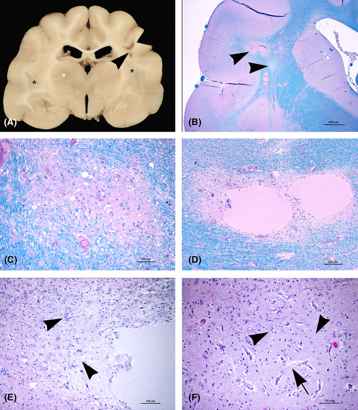
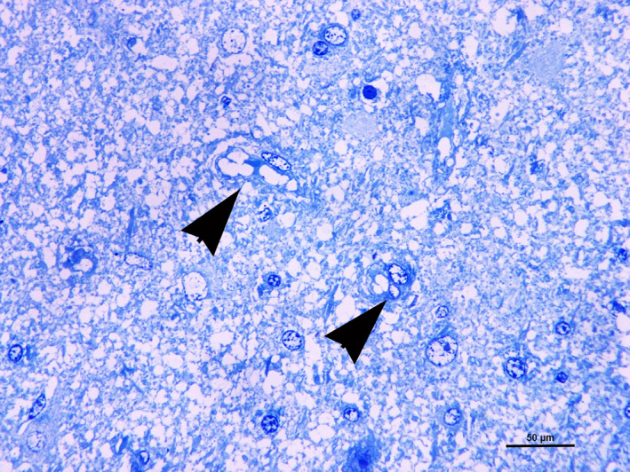
A 2-year-old female cat was referred for progressive neurological signs indicative of involvement of the prosencephalon, cerebellum, and brainstem. Magnetic resonance imaging identified multifocal, bilateral, symmetrical lesions with strong contrast enhancement, affecting multiple areas of the brain. Neuropathology at necropsy showed demyelination, necrotic lesions, spongiosis, and neuropil edema with reactive astrogliosis and neovascularization. Ultrastructural study indicated mitochondrial polymorphism. Genetic investigations outlined 2 polymorphisms within the tRNA-Leu(UUR) gene of mitochondrial DNA. Imaging and neuropathological findings were consistent with selective symmetrical necrotizing encephalopathy, for which genetic investigations support mitochondrial pathogenesis.

Keywords: cat; magnetic resonance imaging; mitochondriopathy; necrotizing encephalopathy; tRNA-Leu(UUR) gene.


Conflict of interest statement

Authors declare no conflict of interest.

Figures

FIGURE 1
FIGURE 1
Transverse magnetic resonance images of the brain at the level of the thalamus in two studies performed 6 months apart. In the initial images (A), there is a diffuse, almost symmetrical, hyperintensity in T2‐weighted images affecting the white matter of both hemispheres and both thalami. These areas are isointense in T1‐weighted images and enhance slightly after contrast medium administration. In the second MRI study (B), the hyperintensity in T2‐weighted images is more pronounced as well as the contrast. MRI, magnetic resonance imaging
FIGURE 2
FIGURE 2
A, Gross lesions. Transverse brain section at level of thalamus. Bilateral areas of discoloration (black asterisks) and cystic lesions (arrowhead) involving the corona radiata. A discoloration area is also present in the thalamus (white asterisk). B‐F, Histological lesions. B, Transverse brain section at the level of nucleus caudatus. The corona radiata shows pale blue areas (arrowheads) consistent with demyelination (LFB‐PAS). C, Detail of (B). Periventricular white matter. Area of LFB discoloration with mild spongiosis (LFB‐PAS). D, Radiate white matter. Necrotic cystic lesions containing pale eosinophilic proteinaceous material (LFB‐PAS). E, Detail of (D), showing a number of reactive astrocytes (gemistocytes) (arrowheads) around the cystic lesion hematoxylin‐eosin (H&E). F, Nucleus caudatus. Increased density of microvessels (neovascularization) (arrow) associated with gliosis (arrowheads) hematoxylin‐eosin (H&E). LFB‐PAS, luxol fast blue‐periodic acid Schiff
FIGURE 3
FIGURE 3
A‐F, Histological lesions. A, Nucleus caudatus. Marked proliferation of endothelial cells in the areas affected by neovascularization (HE). B, Occasional Alzheimer type I cells showing from 3 to 6 small nuclei (arrowheads) (HE). C, Focus of astrocytosis and neovascularization. A number of astrocytes show swollen or lobated nucleus (Alzheimer type II cells) (arrowheads) (HE). D, Cerebellar cortex. Areas of neovascularization in the molecular layer (arrowheads) hematoxylin‐eosin (H&E). E, Cerebellar cortex. In this folium, proliferation of Bergmann's astrocytes is associated to occasional shrunken Purkinje cells consistent with apoptotic bodies hematoxylin‐eosin (H&E). F, Medial vestibular nuclei. Neuronal basophilic inclusions consistent with calcification hematoxylin‐eosin (H&E)
FIGURE 4
FIGURE 4
Cerebral white matter. Diffuse vacuolization of nervous tissue. Two astrocytes show cytoplasm vacuoles (arrowheads) (semithin section, toluidine blue)
FIGURE 5
FIGURE 5
Contextualization of the anticodon stem loop mutation found in this study. On the left panel, the wild‐type 2D structure it tRNA‐Leu with intact alignment of the anticodon stem loop; on the right panel, the 2D structure calculated from our sample. On the bottom, the alignment of the two sequences. In pink is highlighted the mutation as well as its location in the structures. 2D structures were produced with tRNAscan‐SE36

References

    1. Craciun C, Horbin R. Tissue processing schedule for parallel light and electron microscopy. R Microsc Soc Proc. 1989;24:223‐225.
    1. DiMauro S, Schon EA. Mitochondrial disorders in the nervous system. Annu Rev Neurosci. 2008;31:91‐123. - PubMed
    1. Parikh S, Goldstein A, Koenig MK, et al. Diagnosis and management of mitochondrial disease: a consensus statement from the Mitochondrial Medicine Society. Genet Med. 2015;17:689‐701. - PMC - PubMed
    1. Gorman GS, Chinnery PF, DiMauro S, et al. Mitochondrial diseases. Nat Rev Dis Primers. 2016;2:16080. - PubMed
    1. Betts J, Lightowlers RL, Turnbull DM. Neuropathological aspects of mitochondrial DNA disease. Neurochem Res. 2004;29:505‐511. - PubMed

Publication types