

Volume of the right supramarginal gyrus is associated with a maintenance of emotion recognition ability
- PMID: 34293003
- PMCID: PMC8297759
- DOI: 10.1371/journal.pone.0254623
Volume of the right supramarginal gyrus is associated with a maintenance of emotion recognition ability
Abstract
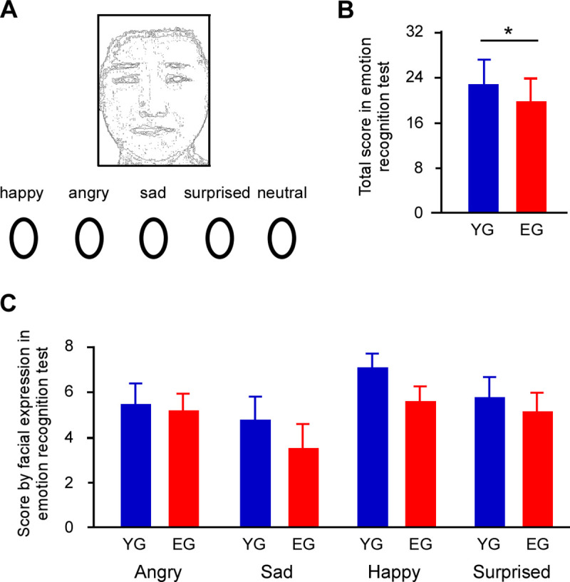
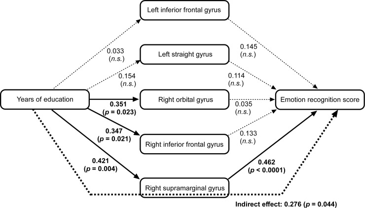
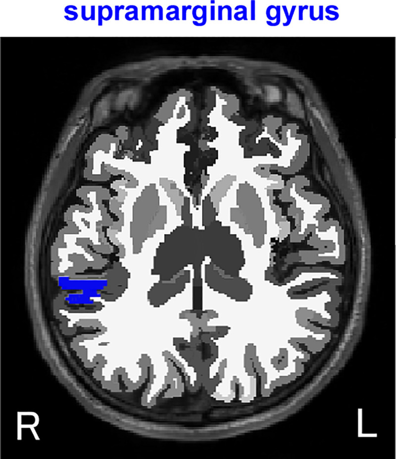
Emotion recognition is known to change with age, but associations between the change and brain atrophy are not well understood. In the current study atrophied brain regions associated with emotion recognition were investigated in elderly and younger participants. Group comparison showed no difference in emotion recognition score, while the score was associated with years of education, not age. We measured the gray matter volume of 18 regions of interest including the bilateral precuneus, supramarginal gyrus, orbital gyrus, straight gyrus, superior temporal sulcus, inferior frontal gyrus, insular cortex, amygdala, and hippocampus, which have been associated with social function and emotion recognition. Brain reductions were observed in elderly group except left inferior frontal gyrus, left straight gyrus, right orbital gyrus, right inferior frontal gyrus, and right supramarginal gyrus. Path analysis was performed using the following variables: age, years of education, emotion recognition score, and the 5 regions that were not different between the groups. The analysis revealed that years of education were associated with volumes of the right orbital gyrus, right inferior frontal gyrus, and right supramarginal gyrus. Furthermore, the right supramarginal gyrus volume was associated with the emotion recognition score. These results suggest that the amount of education received contributes to maintain the right supramarginal gyrus volume, and indirectly affects emotion recognition ability.
Conflict of interest statement
The authors declare no competing interests. The authors also declare that this study was funded by Kao Corporation. The funding body had no influence on design, analysis, and interpretation of the results. Y.M. and H.S. (H.S. is employed by Kao Corporation) were involved in MRI measurements, and were not involved in the subsequent data analysis or interpretation of the results. This does not alter our adherence to PLOS ONE policies on sharing data and materials.
Figures
Similar articles
-
[Resting state fMRI study of emotional network in patients with postconcussion syndrome].Zhonghua Yi Xue Za Zhi. 2017 Jul 4;97(25):1951-1955. doi: 10.3760/cma.j.issn.0376-2491.2017.25.007. Zhonghua Yi Xue Za Zhi. 2017. PMID: 28693073 Chinese.
-
[Mind-regulating and spleen-strengthening needling technique improves abdominal hypersensitivity and emotion by enhancing functional connectivity between hippocampus and brain regions in diarrhea-predominant irritable bowel syndrome patients].Zhen Ci Yan Jiu. 2021 Apr 25;46(4):318-25. doi: 10.13702/j.1000-0607.200569. Zhen Ci Yan Jiu. 2021. PMID: 33931998 Chinese.
-
Neural correlates of successful emotional episodic encoding and retrieval: An SDM meta-analysis of neuroimaging studies.Neuropsychologia. 2020 Jun;143:107495. doi: 10.1016/j.neuropsychologia.2020.107495. Epub 2020 May 13. Neuropsychologia. 2020. PMID: 32416099
-
Cognitive impairment and gray/white matter volume abnormalities in pediatric patients with Turner syndrome presenting with various karyotypes.J Pediatr Endocrinol Metab. 2013;26(11-12):1111-21. doi: 10.1515/jpem-2013-0145. J Pediatr Endocrinol Metab. 2013. PMID: 23846137
-
Facial emotion processing in patients with schizophrenia and their non-psychotic siblings: a functional magnetic resonance imaging study.Schizophr Res. 2012 Feb;134(2-3):143-50. doi: 10.1016/j.schres.2011.10.019. Epub 2011 Nov 22. Schizophr Res. 2012. PMID: 22113155
Cited by
-
Abnormal grey matter structural changes in patients with end-stage kidney disease and mild cognitive impairment: correlations with clinical features.Metab Brain Dis. 2023 Dec;38(8):2817-2829. doi: 10.1007/s11011-023-01293-5. Epub 2023 Sep 30. Metab Brain Dis. 2023. PMID: 37776380 Free PMC article.
-
Aberrant Resting-State Functional Connectivity in MDD and the Antidepressant Treatment Effect-A 6-Month Follow-Up Study.Brain Sci. 2023 Apr 23;13(5):705. doi: 10.3390/brainsci13050705. Brain Sci. 2023. PMID: 37239177 Free PMC article.
-
Increased functional connectivity between brain regions involved in social cognition, emotion and affective-value in psychedelic states induced by N,N-Dimethyltryptamine (DMT).Front Pharmacol. 2024 Oct 30;15:1454628. doi: 10.3389/fphar.2024.1454628. eCollection 2024. Front Pharmacol. 2024. PMID: 39539622 Free PMC article.
-
Resting-state fMRI activation is associated with parent-reported phenotypic features of autism in early adolescence.Front Child Adolesc Psychiatry. 2024 Nov 5;3:1481957. doi: 10.3389/frcha.2024.1481957. eCollection 2024. Front Child Adolesc Psychiatry. 2024. PMID: 39816599 Free PMC article.
-
A dynamic framework of brain functional patterns shaped by spontaneous thoughts beyond the default mode network.Sci Rep. 2025 Aug 4;15(1):28389. doi: 10.1038/s41598-025-10432-0. Sci Rep. 2025. PMID: 40759678 Free PMC article.
References
Publication types
MeSH terms
LinkOut - more resources
Full Text Sources
Medical
