. 2021 Dec 21;42(48):4947-4960.
doi: 10.1093/eurheartj/ehab420.

Effects of canagliflozin on human myocardial redox signalling: clinical implications

Affiliations

Effects of canagliflozin on human myocardial redox signalling: clinical implications

Hidekazu Kondo et al. Eur Heart J. .

Abstract

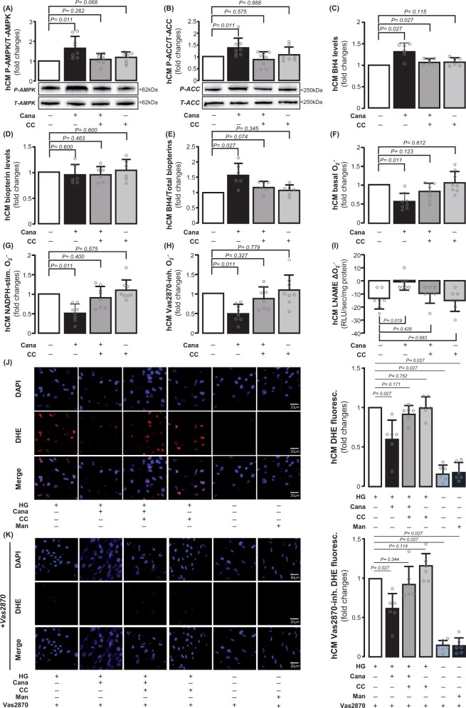
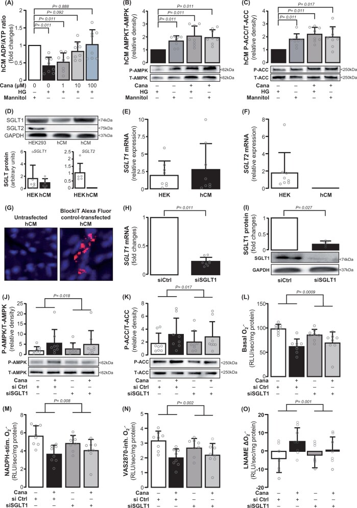
Aims: Recent clinical trials indicate that sodium-glucose cotransporter 2 (SGLT2) inhibitors improve cardiovascular outcomes in heart failure patients, but the underlying mechanisms remain unknown. We explored the direct effects of canagliflozin, an SGLT2 inhibitor with mild SGLT1 inhibitory effects, on myocardial redox signalling in humans.

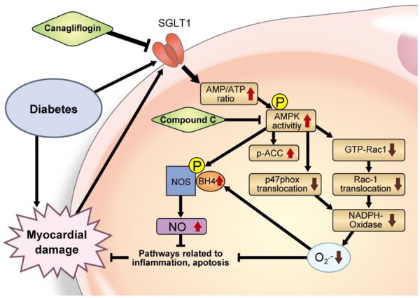
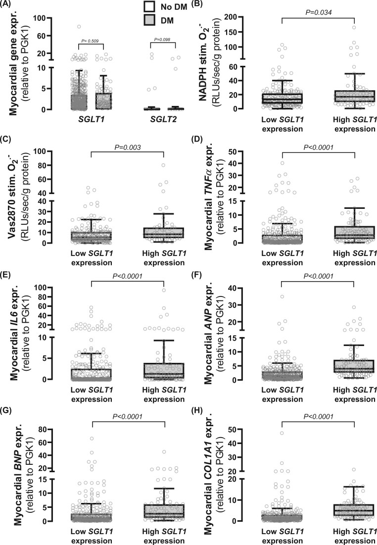
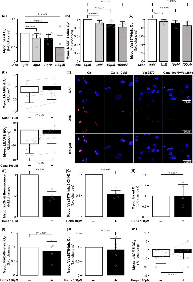
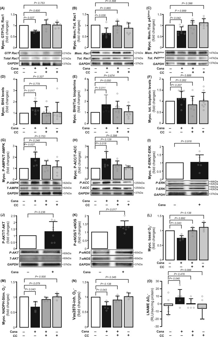
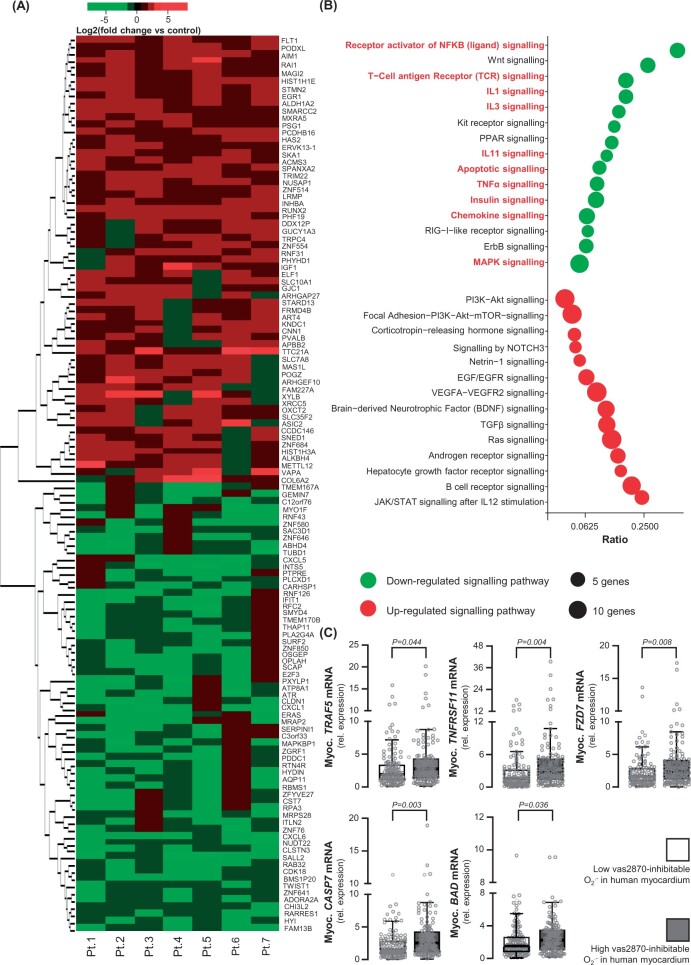
Methods and results: Study 1 included 364 patients undergoing cardiac surgery. Right atrial appendage biopsies were harvested to quantify superoxide (O2.-) sources and the expression of inflammation, fibrosis, and myocardial stretch genes. In Study 2, atrial tissue from 51 patients was used ex vivo to study the direct effects of canagliflozin on NADPH oxidase activity and nitric oxide synthase (NOS) uncoupling. Differentiated H9C2 and primary human cardiomyocytes (hCM) were used to further characterize the underlying mechanisms (Study 3). SGLT1 was abundantly expressed in human atrial tissue and hCM, contrary to SGLT2. Myocardial SGLT1 expression was positively associated with O2.- production and pro-fibrotic, pro-inflammatory, and wall stretch gene expression. Canagliflozin reduced NADPH oxidase activity via AMP kinase (AMPK)/Rac1signalling and improved NOS coupling via increased tetrahydrobiopterin bioavailability ex vivo and in vitro. These were attenuated by knocking down SGLT1 in hCM. Canagliflozin had striking ex vivo transcriptomic effects on myocardial redox signalling, suppressing apoptotic and inflammatory pathways in hCM.

Conclusions: We demonstrate for the first time that canagliflozin suppresses myocardial NADPH oxidase activity and improves NOS coupling via SGLT1/AMPK/Rac1 signalling, leading to global anti-inflammatory and anti-apoptotic effects in the human myocardium. These findings reveal a novel mechanism contributing to the beneficial cardiac effects of canagliflozin.

Keywords: AMPK; Myocardial redox state; NADPH oxidase activity; NOS coupling; SGLT1; SGLT2 inhibitor.


Conflict of interest statement

Conflict of interest: C.A. declared past consultancy agreement with Mitsubishi Tanabe, past grants from Sanofi and Novo Nordisk. C.A. and K.M.C. are founders, shareholders, and directors of Caristo Diagnostics, and C.S. is an employee and shareholder of Caristo Diagnostics. C.A. is the chair of British Atherosclerosis Society. B.C. is the past president of the European Society of Cardiology. The remaining authors have nothing to disclose.

Figures

None
Proposed mechanism of canagliflozin-induced improvement of myocardial redox state. Canagliflozin increases intracellular AMP/ATP ratio through inhibition of SGLT1, which can activate AMPK/NOS signalling and increase NO that suppresses pro-inflammatory signalling. AMPK activation also inhibits activation of Rac1 and membrane translocation of Rac1 and p47phox, which decrease NADPH oxidase activity and superoxide (O2.) production, attenuates inflammatory and apoptotic pathways and increasing the bioavailability of tetrahydrobiopterin (BH4), a key factor for NOS coupling.
Figure 1
Figure 1
Sodium-glucose cotransporter (SGLT)1/2 expression in human atrial myocardium and relations with myocardial redox state and inflammation biomarkers. (A) SGLT1 was abundantly expressed in the human atrial myocardium, contrary to SGLT2. n = 357 [290 non-diabetic patients (no DM); 67 diabetic patients (DM)]. SGLT1 expression was positively correlated with NADPH-stimulated (B) and Vas2870-inhibitable (C) O2. as well as tumour necrosis factor-α (TNFα, D), interleukin-6 (IL6, E), atrial natriuretic peptide (ANP, F), brain natriuretic peptide (BNP, G), and collagen 1A1 (Col1A1, H) expression. P-values by Mann–Whitney U-test for no DM vs. DM (A) and highest quartile of SGLT1 expression vs. rest (B–H). Data are presented as mean ± SD (A) and median [25th–75th percentile] (B–H).
Figure 2
Figure 2
Direct effects of canagliflozin and empagliflozin on human myocardial redox state. Ex vivo canagliflozin (3, 10, and 100 μM) treatment for 1 h reduced basal (A), NADPH-stimulated (B), and Vas2870 inhibitable O2. (C) and increased L-NAME-(delta O2.) (D) dose-dependently in human atrial myocardium. Canagliflozin decreased the intensity of basal and Vas2870 inhibitable 2-hydroxyethidium (2-OH-ethidium) fluorescence (E–G) in human atrial biopsies stained with dihydroethidium (DHE). Empagliflozin had non-significant direct effect on either of these measures (H–K). n = 5–7 in panels A–L. Data are presented as mean ± SD. P-values are calculated by Wilcoxon signed-rank test (A–C, F–J) and paired t-test (D, K).
Figure 3
Figure 3
Effects of canagliflozin on myocardial NADPH oxidase activity and nitric oxide synthase (NOS) coupling status in the human atrial myocardium. Canagliflozin (10 μM for 1 h) inhibited GTP activation (A) and membrane translocation (B) of Rac1, as well as the membrane translocation of p47phox (C). Canagliflozin increased myocardial BH4 but not total biopterin content (D–F). Canagliflozin induced AMPK Thr172 phosphorylation (G) and downstream acetyl-coA carboxylase (ACC) Ser79 phosphorylation (H). These were prevented by the AMPK inhibitor, compound C (CC) (G and H). Canagliflozin did not affect ERK or AKT phosphorylation (I and J). Canagliflozin induced NOS Ser1177 phosphorylation (K). Compound C prevented the effects of canagliflozin on Rac1 activation, BH4 bioavailability (A-F), O2. generation (L–N), and NOS coupling (O). n = 5–8 pairs in panels A–O. Data are presented as mean ± SD. P-values are calculated by Wilcoxon signed-rank test (A–N) and paired t-test (O).
Figure 4
Figure 4
Direct effects of canagliflozin on human cardiomyocytes (hCM). Canagliflozin (10 μΜ) induced phosphorylation of AMPK and acetyl-coA carboxylase (ACC) in high glucose (HG)-treated human cardiomyocytes (A and B, n = 8) and increased BH4 levels without affecting total biopterin levels (C–E, n = 8). Canagliflozin decreased basal, NADPH-stimulated and Vas2870-inhibitable O2. and increased the value of L-NAME delta(O2.) in human cardiomyocytes (F–I, n = 7). Dihydroethidium (DHE) staining combined with Vas2870 confirmed these (J–L, n = 8). Data are presented as mean ± SD. P-values are calculated by Wilcoxon signed-rank test (A–H, J, K) and paired t-test (I).
Figure 5
Figure 5
SGLT1 mediates the effects of canagliflozin on myocardial redox state. Canagliflozin increased ADP/ATP dose-dependently in human cardiomyocytes, while glucose deprivation from the culture medium (NG) induced similar changes in ADP/ATP ratio (A, n = 8). There were no differences in AMPK and ACC phosphorylation among the NG-incubated cells with or without canagliflozin (10μΜ), and high glucose (HG)-incubated cells with canagliflozin (panels B and C, n = 8). SGLT1 expression was detected, while SGLT2 was not detected in human cardiomyocytes (D–F, n = 8). SGLT1 was knocked down using siRNA (transfection low toxicity and efficiency was confirmed by transfecting BlockiT Alexa Fluor Red Fluorescent control, G), resulting into ∼76% down-regulation of SGLT1 mRNA (panel H, n = 6), and ∼85% protein down-regulation (panel I, n = 6). Canagliflozin-induced AMPK and ACC phosphorylation was attenuated in SGLT1 knocked down human cardiomyocytes compared with siRNA ctrl human cardiomyocytes (J and K, n = 8). SGLT1 deletion diminished canagliflozin-induced decrease of basal (L), NADPH-stimulated (M), and Vas2870-inhibitable O2. production (N) and increased L-NAME delta O2. (O). n = 8 in L–O. Data are presented as mean ± SD. P-values are calculated by Wilcoxon signed-rank test (A–C, H, I). Comparisons in canagliflozin responses between siControl and siSGLT1 cells were performed with two-way ANOVA with treatment (canagliflozin) × cell type (siControl or siSGLT1) interaction (J–O).
Figure 6
Figure 6
Canagliflozin has a global anti-inflammatory and anti-apoptotic effect on human cardiomyocytes. In this experiment, human cardiomyocytes were cultured in high-glucose medium (25 mM) for 72 h and then treated with canagliflozin (10 μM) or DMSO for 24 h. Heat map of 127 down- or up-regulated genes by canagliflozin (fold change >1.5 or `−1.5, P < 0.05) in canagliflozin-treated human cardiomyocytes from n = 7 patients (A). NFkB, Wnt, IL1, IL3, TNFα, chemokine, MAPK pathways, and apoptotic pathways (highlighted by red font) were down-regulated by canagliflozin (more than 50% of pathway genes down-regulated by canagliflozin, B). TNFRSF11, TRAF5, FZD7, CASP7, and BAD were the most down-regulated individual genes upon canagliflozin treatment, implicated in NFkB, TNFα, and apoptosis pathways. The mRNA expression of these genes was positively correlated with myocardial NADPH oxidase activity (i.e. high Vas2870-inhibitable signal) in 240 atrial biopsies from Study 1 (C). Data are presented as median [25th–75th percentile]. P-values are calculated by Mann–Whitney U-test for high (above median) vs. low (below median).

Comment in

References

    1. Ferrannini E, Solini A. SGLT2 inhibition in diabetes mellitus: rationale and clinical prospects. Nat Rev Endocrinol 2012;8:495—502. - PubMed
    1. Kurosaki E, Ogasawara H. Ipragliflozin and other sodium-glucose cotransporter-2 (SGLT2) inhibitors in the treatment of type 2 diabetes: preclinical and clinical data. Pharmacol Ther 2013;139:51—59. - PubMed
    1. Danne T, Biester T, Kordonouri O. Combined SGLT1 and SGLT2 inhibitors and their role in diabetes care. Diabetes Technol Ther 2018;20:S269—S277. - PubMed
    1. Zinman B, Wanner C, Lachin JM, Fitchett D, Bluhmki E, Hantel S, Mattheus M, Devins T, Johansen OE, Woerle HJ, Broedl UC, Inzucchi SE; EMPA-REG OUTCOME Investigators. Empagliflozin, cardiovascular outcomes, and mortality in type 2 diabetes. N Engl J Med 2015;373:2117—2128. - PubMed
    1. Neal B, Perkovic V, Mahaffey KW, de Zeeuw D, Fulcher G, Erondu N, Shaw W, Law G, Desai M, Matthews DR; CANVAS Program Collaborative Group. Canagliflozin and cardiovascular and renal events in type 2 diabetes. N Engl J Med 2017;377:644—657. - PubMed

Publication types

MeSH terms

Substances