Review
. 2021 Aug 18;109(16):2519-2534.
doi: 10.1016/j.neuron.2021.06.026. Epub 2021 Jul 21.

Spontaneous activity in developing thalamic and cortical sensory networks

Affiliations
Review

Spontaneous activity in developing thalamic and cortical sensory networks

Francisco J Martini et al. Neuron. .

Abstract

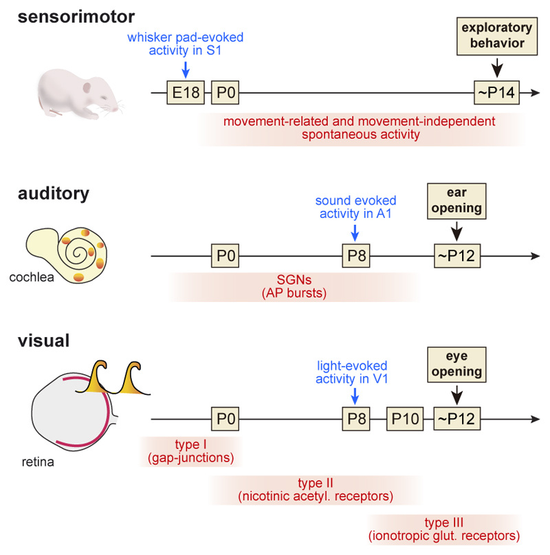
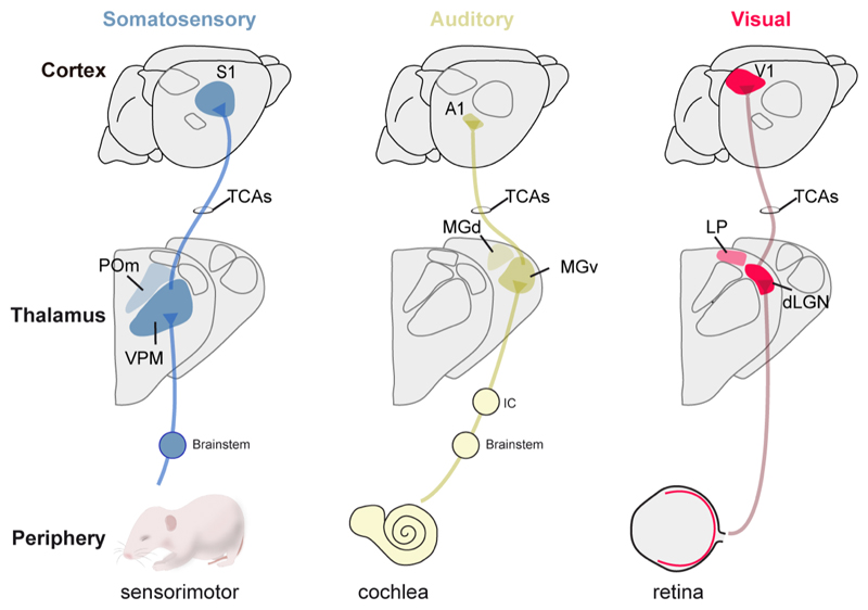
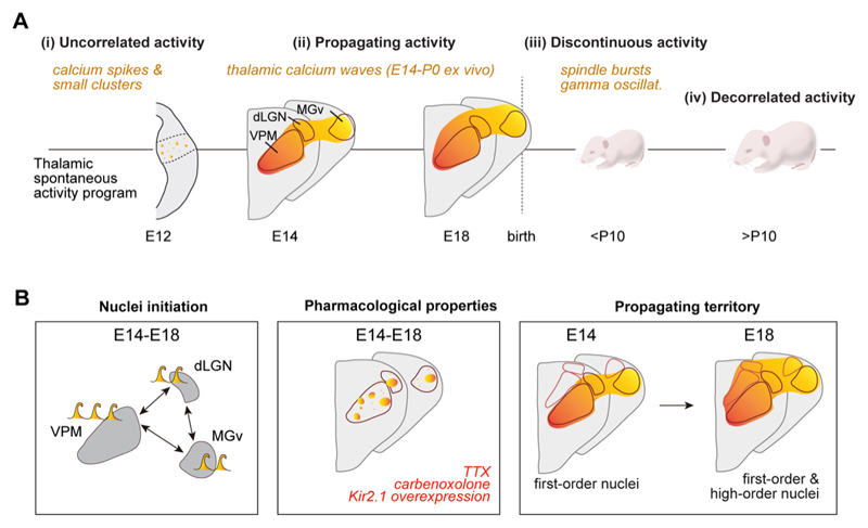
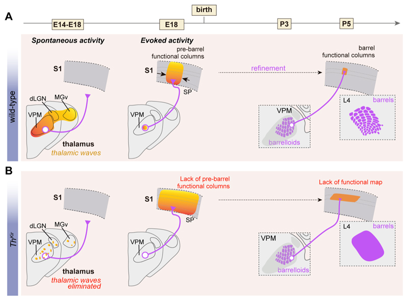
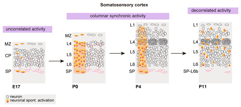
Developing sensory circuits exhibit different patterns of spontaneous activity, patterns that are related to the construction and refinement of functional networks. During the development of different sensory modalities, spontaneous activity originates in the immature peripheral sensory structures and in the higher-order central structures, such as the thalamus and cortex. Certainly, the perinatal thalamus exhibits spontaneous calcium waves, a pattern of activity that is fundamental for the formation of sensory maps and for circuit plasticity. Here, we review our current understanding of the maturation of early (including embryonic) patterns of spontaneous activity and their influence on the assembly of thalamic and cortical sensory networks. Overall, the data currently available suggest similarities between the developmental trajectory of brain activity in experimental models and humans, which in the future may help to improve the early diagnosis of developmental disorders.

Keywords: developing brain; sensory systems; spontaneous activity.


Conflict of interest statement

Declaration of interests The authors declare no competing interests.

Figures

Figure 1
Figure 1. Early manifestations of peripheral spontaneous and evoked activity in developing sensory systems in the mouse.
Developmental progression of peripheral spontaneous (in red) and evoked (in blue) activity in sensorimotor, auditory and visual systems. In the somatosensory system the peripheral activity may be mediated by sensory spontaneous activity or through a sensory-motor feedback. In the auditory system the developing cochlea generates spontaneous activity that is transmitted to SGNs as action potential (AP) bursts. In the visual system the developing retina generates spontaneous waves of activity with a defined developmental profile that is transmitted to the visual thalamus and cortex. E= embryonic age; P= postnatal age; SGN=spiral ganglion neurons.
Figure 2
Figure 2. Connectivity from the sensory periphery to the thalamus and cortex.
Schema representing the three major inputs from the peripheral organs to thalamic sensory nuclei and primary sensory cortical areas. First-order sensory nuclei (VPM, dLGN and MGv) receive direct input from peripheral pathways. High-order nuclei (POm, LP and MGd) receive feedback input from sensory cortices. TCAs= thalamocortical axons, POm= postero-medial nucleus, VPM= ventral posteromedial nucleus, MGd= dorsal medial geniculate, MGv= ventral medial geniculate nucleus, LP= lateral posterior nucleus, dLGN= dorso-lateral geniculate nucleus.
Figure 3
Figure 3. Developmental properties of thalamic spontaneous activity.
(A) The pattern of thalamic spontaneous activity is modified during embryonic and early postnatal development. (B) Properties of embryonic thalamic waves from ex vivo experiments. Left: spontaneous waves can be originated in any of the principal nuclei, but most frequently in the VPM. Middle: initially, waves spread to cover the three principal first-order nuclei while at perinatal stages the activity extends to high-order nuclei. Right: voltage-dependent Na-channel blocker TTX, gap-junction blocker carbonoxolone, or overexpression of the inward rectifier Kir2.1 channel blocks correlated spontaneous activity and only asynchronous activity and small clusters are left in the thalamus. Color gradient represents time course of activation. E= embryonic age; P= postnatal age.
Figure 4
Figure 4. Role of thalamic spontaneous activity on the formation of cortical maps.
(A) Under normal conditions, embryonic thalamic waves emerge around E14 at the time thalamocortical axons (purple) navigate to the cortex. Small VPM stimulations before and soon after birth reveal that S1 cortical responses are fairly columnar. Around P4, similar VPM stimulations trigger columnar responses restricted to L4 as thalamocortical axons already target this layer. (B) In the absence of thalamic waves, with only sparse and asynchronous events in the thalamus, thalamocortical axonal navigation appears spared, however restricted thalamic VPM activation elicits a widespread cortical activity at early stages. At later stages, VPM activation leads to a L4-restricted but not columnar cortical response. Barrels do not form in Thkir mice. Color gradient represents time course of activation. E= embryonic age; P= postnatal age, VPM= ventral posteromedial nucleus, MGv= ventral medial geniculate nucleus, dLGN= dorso-lateral geniculate nucleus, SP= subplate.
Figure 5
Figure 5. Developmental progression of cortical spontaneous activity.
At embryonic stages, cortical spontaneous activity is uncorrelated and restricted to single cells or small clusters. From birth, cortical spontaneous activity is detected in a columnar fashion that at P4 coincides with the dimension of a barrel. Active whisking is associated with the decorrelation and sparsification of cortical activity. E= embryonic age; P= postnatal age, MZ= marginal zone, CP= cortical plate, SP= subplate.

References

    1. Ackman JB, Burbridge TJ, Crair MC. Retinal waves coordinate patterned activity throughout the developing visual system. Nature. 2012;490:219–225. doi: 10.1038/nature11529. - DOI - PMC - PubMed
    1. Ackman JB, Crair MC. Role of emergent neural activity in visual map development. Current Opinion in Neurobiology. 2014;24:166–175. doi: 10.1016/j.conb.2013.11.011. - DOI - PMC - PubMed
    1. Akerman CJ, Smyth D, Thompson ID. Visual experience before eye-opening and the development of the retinogeniculate pathway. Neuron. 2002;36:869–879. doi: 10.1016/s0896-6273(02)01010-3. - DOI - PubMed
    1. Allene C, Cattani A, Ackman JB, Bonifazi P, Aniksztejn L, Ben-Ari Y, Cossart R. Sequential Generation of Two Distinct Synapse-Driven Network Patterns in Developing Neocortex. Journal of Neuroscience. 2008;28:12851–12863. doi: 10.1523/JNEUROSCI.3733-08.2008. - DOI - PMC - PubMed
    1. An S, Kilb W, Luhmann HJ. Sensory-evoked and spontaneous gamma and spindle bursts in neonatal rat motor cortex. Journal of Neuroscience. 2014;34:10870–10883. doi: 10.1523/JNEUROSCI.4539-13.2014. - DOI - PMC - PubMed

Publication types

LinkOut - more resources