. 2021 Aug 3;66(16):10.1088/1361-6560/ac16ea.
doi: 10.1088/1361-6560/ac16ea.

HEDOS-a computational tool to assess radiation dose to circulating blood cells during external beam radiotherapy based on whole-body blood flow simulations

Affiliations

HEDOS-a computational tool to assess radiation dose to circulating blood cells during external beam radiotherapy based on whole-body blood flow simulations

Jungwook Shin et al. Phys Med Biol. .

Abstract

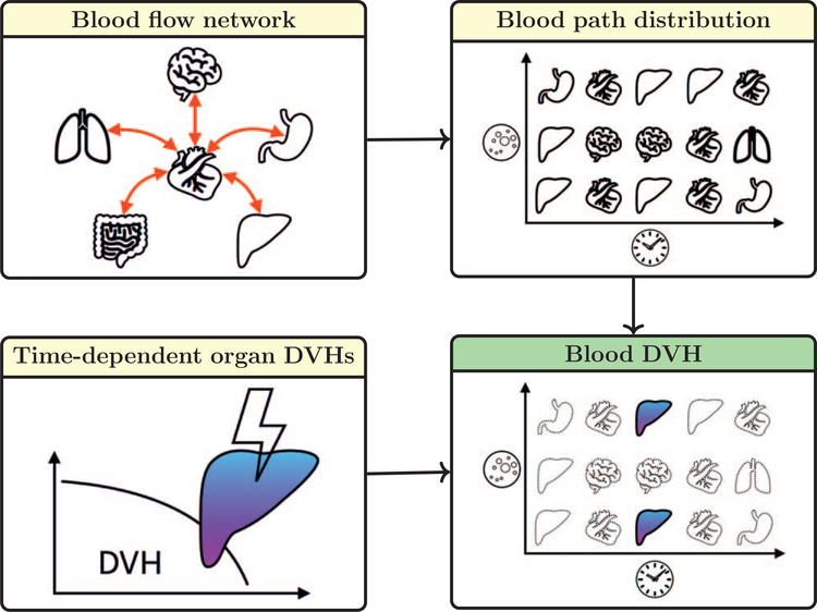
We have developed a time-dependent computational framework, hematological dose (HEDOS), to estimate dose to circulating blood cells from radiation therapy treatment fields for any treatment site. Two independent dynamic models were implemented in HEDOS: one describing the spatiotemporal distribution of blood particles (BPs) in organs and the second describing the time-dependent radiation field delivery. A whole-body blood flow network based on blood volumes and flow rates from ICRP Publication 89 was simulated to produce the spatiotemporal distribution of BPs in organs across the entire body using a discrete-time Markov process. Constant or time-varying transition probabilities were applied and their impact on transition time was investigated. The impact of treatment time and anatomical site were investigated using imaging data and dose distributions from a liver cancer and a brain cancer patient. The simulations revealed different dose levels to the circulating blood for brain irradiation compared to liver irradiation even for similar field sizes due to the different blood flow properties of the two organs. The volume of blood receiving any dose (V>0 Gy) after a single radiation fraction increases from 1.2% for a 1 s delivery time to 20.9% for 120 s delivery time for the brain cancer treatment, and from 10% (1 s) to 48.7% (120 s) for a liver cancer treatment. Other measures of the low-dose bath to the circulating blood such as the dose to small volumes of blood (D2%) decreases with longer delivery time. Furthermore, we demonstrate that the blood dose-volume histogram is highly sensitive to changes in the treatment time, indicating that dynamic modeling of blood flow and radiation fields is necessary to evaluate dose to circulating blood cells for the assessment of radiation-induced lymphopenia. HEDOS is publicly available and allows for the estimation of patient-specific dose to circulating blood cells based on organ DVHs, thus enabling the study of the impact of different treatment plans, dose rates, and fractionation schemes.

Keywords: discrete-time Markov process; lymphopenia; radiation dose to lymphocytes; radiotherapy; whole-body blood flow.


Figures

Figure 1.
Figure 1.
HEDOS: diagram of the modularized components and their processes to compute BP doses. First, a whole-body organ network connected by transition probabilities is constructed. Using a discrete-time Markov process, a spatiotemporal BP path distribution is generated. Next, the accumulation of absorbed dose is performed for the BPs which reside in the irradiated organs during the period that a radiation field is delivered.
Figure 2.
Figure 2.
(A) Diagram of the blood flow through 28 organs according to ICRP Publication 89 (Valentin 2002). (B) Transition probability matrix per second based on ICRP reference data. Each element corresponds to the transition probability of a BP from a given row to a given column. For all simulations in this study, we use the reference male total blood volume of 5.3l and cardiac output of 6.3lmin−1, according to ICRP 89.
Figure 3.
Figure 3.
(A) Initial age distribution from a Weibull distribution whose к and λ values are 2 s and 19.2 s, respectively. (B) Exit probabilities for constant and time-dependent transition probabilities. Distributions of exiting and remaining BPs using time-dependent transition probabilities (C) and constant exiting probability (D).
Figure 4.
Figure 4.
Diagram explaining how a BP receives absorbed dose by using a blood path with a time step (dt). Two fields (DVH0 and DVH1 ) were delivered sequentially with a break with delivery times of T0 and T1. At each time step in the irradiated organ, an absorbed dose value is sampled from dose rate functions, say f0 and f1. The absorbed dose of the BP (d) is the sum of doses (d0 and d1 ) from two fields. A partial delivery (less than one time-step) is also taken into account.
Figure 5.
Figure 5.
DVHs for the entire brain and liver per fraction for a brain tumor treatment with six fields (A) and a liver tumor treatment with two fields (B), respectively.
Figure 6.
Figure 6.
A) Six regions of the ICRP Publication 145 mesh-based adult male reference phantom (Kim et al 2020) defined by a pair of planes based on our institution’s CT imaging protocol. The relative volumes of muscle, fat, skin, large vasculature and bone of each region were calculated to distribute dose to un-contoured areas. (B) Relative volume of tissues of interest in these six regions as percentage of that compartment in the entire body.
Figure 7.
Figure 7.
Spatiotemporal blood path distribution of 105 BPs for 10 min. A pixel has an organ index indicating the position and time of a BP. Colors are used to distinguish compartments.
Figure 8.
Figure 8.
Comparisons of averaged blood volumes over 10 min between constant transition probability and time-dependent transition probability (Weibull). The green bars are the ICRP reference values.
Figure 9.
Figure 9.
Transition time distributions for brain (A) and liver (B) with the two transition probability models. Mean transition time (MTT) and mean return time (MRT) for all organs with transition probability models are shown in (C) for constant and (D) for Weibull, respectively. MRT of gonads for Weibull (k=2) was not defined because not a single BP returned within 10 min.
Figure 10.
Figure 10.
The impact of beam delivery time on bDVHs and their metrics, such as V>0 Gy, V0.05 Gy,,and D2%, for brain irradiation (A)-(F) and liver irradiation (G)–(L): (A)–(D) blood dose-volume histograms (bDVHs) for brain irradiation, (G)–(J) for liver irradiation. The bDVH metrics, V>0 Gy,, V0.05 Gy and D2%, are plotted as a function of the beam delivery time.
Figure 11.
Figure 11.
The impact of beam delivery breaks on bDVHs and their metrics for brain (A)–(F) and liver irradiation (G)–(L): bDVHs as a function of beam interval between fields for brain (A)–(D) and liver irradiation (G)–(J). The bDVH metrics, V>0 Gy,V0.05 Gy and D2%, are plotted asa function of the beam interval time.
Figure 12.
Figure 12.
bDVH as a function of fraction size for brain irradiation (A) and liver irradiation (D). The bDVH metrics, V>0 Gy,V0.05 Gy and D2%, are plotted as a function of fraction size for brain (B) and (C) and liver irradiation (E )and (F).

References

    1. Balmanoukian A, Ye X, Herman J, Laheru D and Grossman SA 2012. The association between treatment-related lymphopenia and survival in newly diagnosed patients with resected adenocarcinoma of the pancreas Cancer Invest. 30 571–6 - PMC - PubMed
    1. Basler L, Andratschke N, Ehrbar S, Guckenberger M and Tanadini-Lang S 2018. Modelling the immunosuppressive effect of liver SBRT by simulating the dose to circulating lymphocytes: anin-silico planning study Radiat. Oncol. 13 10. - PMC - PubMed
    1. Borgmann K et al. 2008. Individual radiosensitivity measured with lymphocytes may predict the risk of acute reaction after radiotherapy Int.J. Radiat. Oncol. 71 256–64 - PubMed
    1. Byun HK,Kim N, Park S and Seong J 2019. Acute severe lymphopenia by radiotherapy is associated with reduced overall survival in hepatocellular carcinoma Strahlenther. Onkol. 195 1007–17 - PubMed
    1. Catalano OA, Singh AH, Uppot RN, Hahn PF, Ferrone CR and Sahani DV 2008. Vascular and biliary variants in the liver:implications for liver surgery RadioGraphics 28 359–78 - PubMed

Publication types