. 2021 Jul 22;12(1):416.
doi: 10.1186/s13287-021-02492-6.

hucMSC-derived exosomes attenuate colitis by regulating macrophage pyroptosis via the miR-378a-5p/NLRP3 axis

Affiliations

hucMSC-derived exosomes attenuate colitis by regulating macrophage pyroptosis via the miR-378a-5p/NLRP3 axis

Xiu Cai et al. Stem Cell Res Ther. .

Abstract

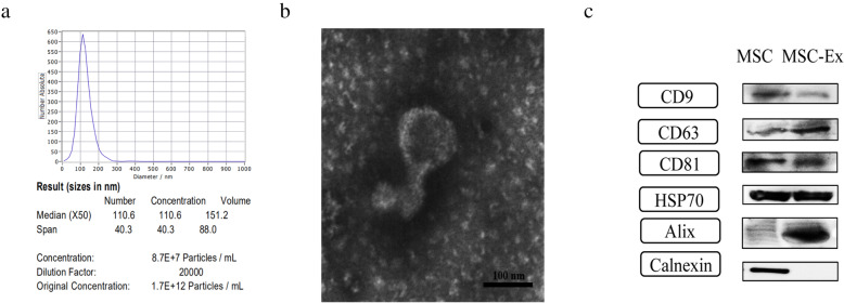
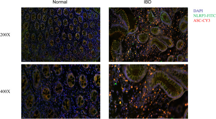
Background: Human umbilical cord mesenchymal stem cell (hucMSC)-derived exosomes are recognized as novel cell-free therapeutic agents for inflammatory bowel disease (IBD), a condition caused by dysregulated intestinal mucosal immunity. In this event, macrophage pyroptosis, a process of cell death following the activation of NLRP3 (NOD-like receptor family, pyrin domain-containing 3) inflammasomes, is believed to partially account for inflammatory reactions. However, the role of macrophage pyroptosis in the process of hucMSC-derived exosomes alleviating colitis remains unknown. This study aimed at exploring the therapeutic effect and mechanism of hucMSC-derived exosomes on colitis repair.

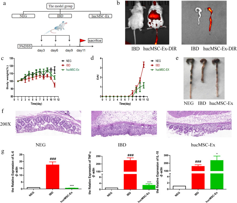
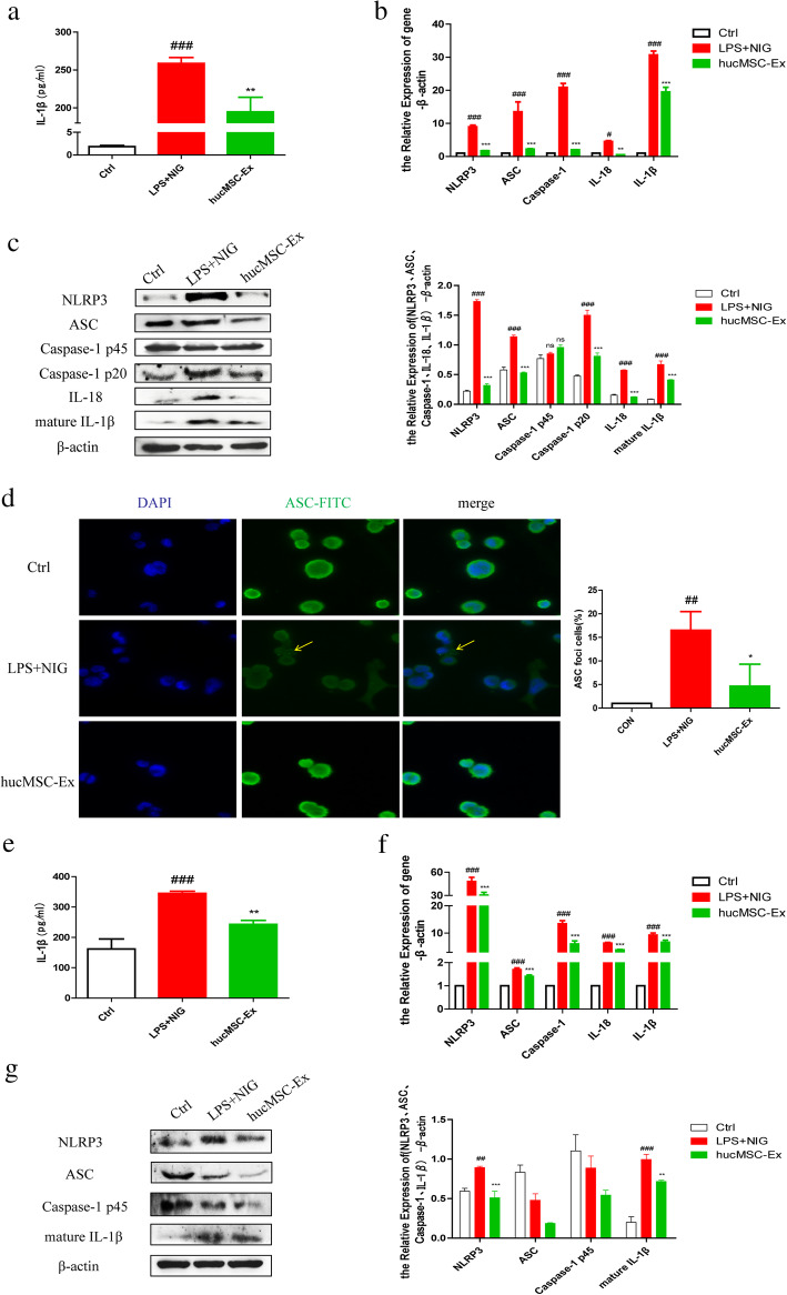
Methods: In vivo, we used BALB/c mice to establish a dextran sulfate sodium (DSS)-induced colitis model and administrated hucMSC-derived exosomes intravenously to estimate its curative effect. Human myeloid leukemia mononuclear (THP-1) cells and mouse peritoneal macrophages (MPMs) were stimulated with lipopolysaccharides (LPS) and Nigericin to activate NLRP3 inflammasomes, which simulated an inflammation environment in vitro. A microRNA mimic was used to verify the role of miR-378a-5p/NLRP3 axis in the colitis repair.

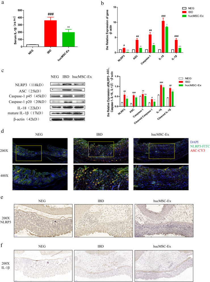
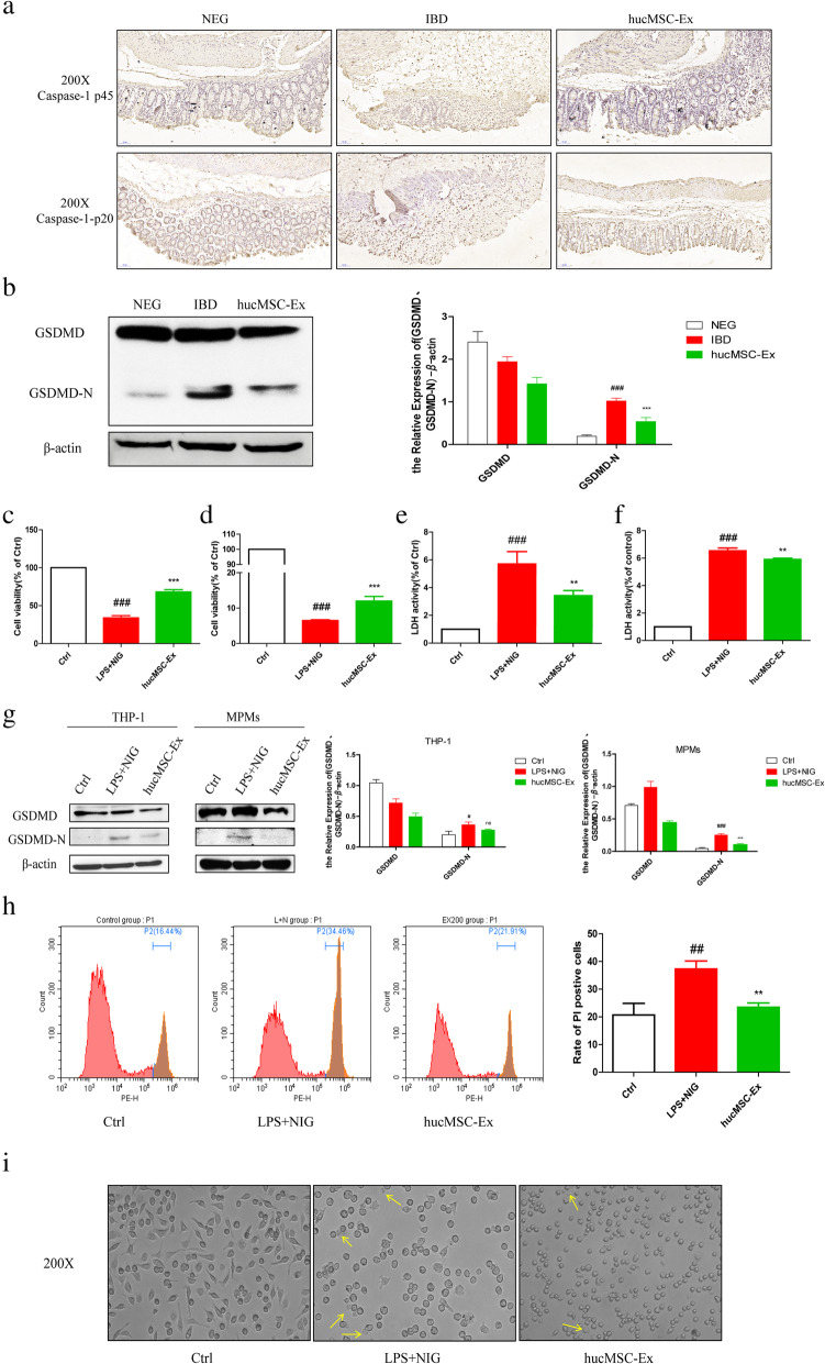
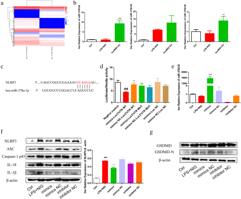
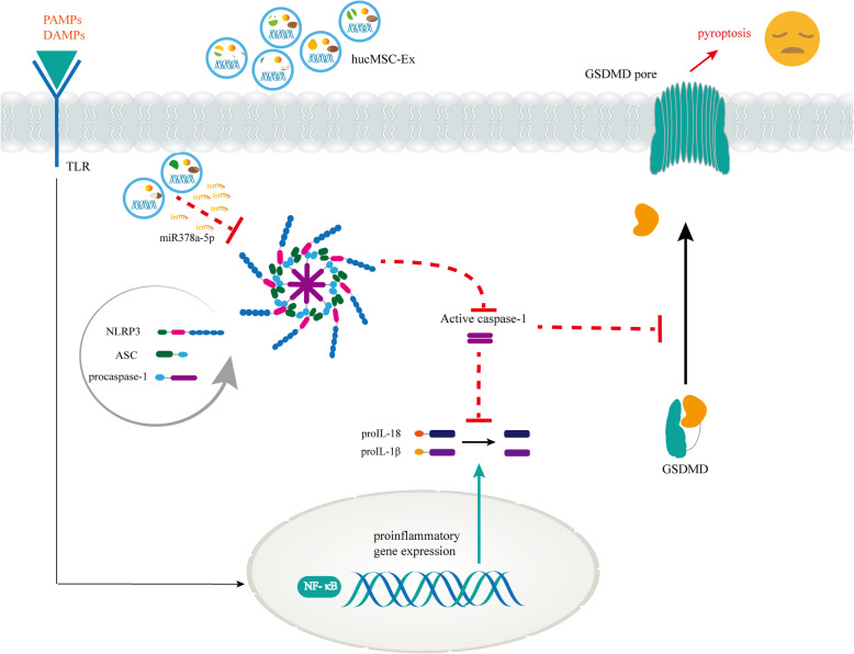
Results: hucMSC-derived exosomes inhibited the activation of NLRP3 inflammasomes in the mouse colon. The secretion of interleukin (IL)-18, IL-1β, and Caspase-1 cleavage was suppressed, resulting in reduced cell pyroptosis. The same outcome was observed in the in vitro cell experiments, where the co-culture of THP-1 cells and MPMs with hucMSC-derived exosomes caused decreased expression of NLRP3 inflammasomes and increased cell survival. Furthermore, miR-378a-5p was highly expressed in hucMSC-derived exosomes and played a vital function in colitis repair.

Conclusion: hucMSC-derived exosomes carrying miR-378a-5p inhibited NLRP3 inflammasomes and abrogated cell pyroptosis to protect against DSS-induced colitis.

Keywords: IBD; Macrophage; NLRP3; Pyroptosis; hucMSC-derived exosomes; miR-378a-5p.


Conflict of interest statement

The authors declare that they have no competing interests.

Figures

Fig. 1
Fig. 1
Identification of hucMSC-derived exosomes. a NanoSight Nanoparticle Tracking Analyzer detection of hucMSC-derived exosome diameters. b Transmission electron microscope observation of hucMSC-derived exosome morphology. c Western blot analysis of hucMSC-derived exosome protein markers
Fig. 2
Fig. 2
hucMSC-derived exosomes alleviate DSS-induced mice colitis. a Colitis model established. The model contained the control (Neg), IBD, and hucMSC-Ex groups. 3% DSS was given at day 1 to mice in the IBD group and hucMSC-Ex group. Mice in the hucMSC-Ex group were given hucMSC-derived exosomes iv. at days 3, 6, and 9. b Fluorescence distribution of DIR-labeled hucMSC-derived exosomes in mouse colon after injection. c The mouse body weight loss. d DAI index of mice. e Mouse colon appearance. f H&E staining of mouse colon (200×, scale bar = 50 μm). g QRT-PCR analysis of the expression of inflammatory cytokines (IL-6, TNF-α, IL-10) in mouse colon tissues. #P < 0.05, ##P < 0.01, ###P < 0.001 vs Ctrl by ANOVA; *P < 0.05, **P < 0.01, ***P < 0.001 vs LPS + NIG by ANOVA
Fig. 3
Fig. 3
hucMSC-derived exosomes protect against DSS-induced colitis via inhibiting NLRP3 inflammasomes. a The quantitative analysis of mouse serum IL-1β (pg ml−1) by ELISA. b QRT-PCR analysis of the mRNA expression level of NLRP3 inflammasome-related molecules in mouse colon tissues. c Western blot analysis of the protein expression level of NLRP3 inflammasome-related molecules in mouse colon tissues and its grayscale scanning analysis. d Representative images of IF staining for NLRP3 and ASC on sections of mouse colon tissues (200×, scale bar = 50 μm; 400×, scale bar = 20 μm). e IHC analysis of NLRP3 expression in mouse colon tissues (200×, scale bar = 50 μm). f IHC analysis of IL-1β expression in the mouse colon tissues (200×, scale bar = 50 μm). #P < 0.05, ##P < 0.01, ###P < 0.001 vs Ctrl by ANOVA; *P < 0.05, **P < 0.01, ***P < 0.001 vs LPS + NIG by ANOVA
Fig. 4
Fig. 4
Increased expression of NLRP3 inflammasomes in IBD patients’ colon tissues compared with healthy controls. Representative images of IF staining for NLRP3 and ASC (200×, scale bar = 50 μm; 400×, scale bar = 20 μm)
Fig. 5
Fig. 5
hucMSC-derived exosomes decrease the activation of NLRP3 inflammasomes in macrophages. a The quantitative analysis of IL-1β (pg ml−1) in THP-1 cell supernatant by ELISA. b QRT-PCR analysis of the mRNA expression level of NLRP3 inflammasome-related molecules in THP-1 cells. c Western blot analysis of the protein expression level of NLRP3 inflammasome-related molecules in THP-1 cell lysate and grayscale scanning analysis. d IF analysis of ASC oligomerization in THP-1 cells (scale bar = 50 μm) and the statistical analysis of the percentage of ASC foci cells. e The quantitative analysis of IL-1β (pg ml−1) in MPM supernatant by ELISA. f QRT-PCR analysis of the mRNA expression level of NLRP3 inflammasome-related molecules in MPMs. g Western blot analysis of the protein expression level of NLRP3 inflammasome-related molecules in MPMs lysate and grayscale scanning analysis. #P < 0.05, ##P < 0.01, ###P < 0.001 vs Ctrl by ANOVA; *P < 0.05, **P < 0.01, ***P < 0.001 vs LPS + NIG by ANOVA
Fig. 6
Fig. 6
hucMSC-derived exosomes reduce cell pyroptosis by decreasing the activation of NLRP3 inflammasomes both in vivo and in vitro. a IHC analysis of Caspase-1 p45 and Caspase-1 p20 expression in mouse colon tissues (200×, scale bar = 50 μm). b Western blot analysis of GSDMD protein levels in mouse colon tissues and the grayscale scanning analysis. c CCK8 assay analysis of THP-1 cell viability. d CCK8 assay analysis of MPM viability. e LDH activity analysis of THP-1 cell supernatant. f LDH activity analysis of MPM supernatant. g Identification of PI-positive THP-1 cells by flow cytometry. h Western blot analysis of the protein expression level of GSDMD and cleaved GSDMD fragment in both THP-1 cell lysate and MPM lysate and the grayscale scanning analysis. i Imaging assay of pyroptosis in MPMs treated as indicated. #P < 0.05, ##P < 0.01, ###P < 0.001 vs Ctrl by ANOVA; *P < 0.05, **P < 0.01, ***P < 0.001 vs LPS + NIG by ANOVA
Fig. 7
Fig. 7
miR-378a-5p is a key molecule in the inhibitory effect of hucMSC-exosomes on NLRP3 inflammasome activation. a Sequence results of miRNAs in hucMSC-derived exosomes and HFL1-derived exosomes. b QRT-PCR analysis of miR-378a-5p expression levels in THP-1 cells treated as indicated. c Binding sites between NLRP3 and miR-378a-5p. d Dual-luciferase reporter gene detection of the targeting relationship between NLRP3 and miR-378a-5p. e QRT-PCR analysis of the miR-378a-5p in THP-1 cells after transfection. f Western blot analysis of the protein expression level of NLRP3 inflammasome-related molecules in THP-1 cell lysate after transfection and the grayscale scanning analysis of NLRP3. g Western blot analysis of the protein expression level of GSDMD and cleaved GSDMD fragment in THP-1 cell lysate after transfection. #P < 0.05, ##P < 0.01, ###P < 0.001 vs Ctrl by ANOVA; *P < 0.05, **P < 0.01, ***P < 0.001 vs LPS + NIG (mimics Luc3′UTR WT) by ANOVA
Fig. 8
Fig. 8
The mechanistic model of miR-378a-5p in hucMSC-derived exosome-mediated NLRP3 inflammasome activation and cell pyroptosis

References

    1. Marion-Letellier R, Savoye G, Ghosh S. IBD: In food we trust. J Crohns Colitis. 2016;10(11):1351–1361. doi: 10.1093/ecco-jcc/jjw106. - DOI - PubMed
    1. de Souza HS, Fiocchi C. Immunopathogenesis of IBD: current state of the art. Nat Rev Gastroenterol Hepatol. 2016;13(1):13–27. doi: 10.1038/nrgastro.2015.186. - DOI - PubMed
    1. Kaplan GG. The global burden of IBD: from 2015 to 2025. Nat Rev Gastroenterol Hepatol. 2015;12(12):720–727. doi: 10.1038/nrgastro.2015.150. - DOI - PubMed
    1. Lissner D, Schumann M, Batra A, Kredel LI, Kuhl AA, Erben U, et al. Monocyte and M1 macrophage-induced barrier defect contributes to chronic intestinal inflammation in IBD. Inflamm Bowel Dis. 2015;21(6):1297–1305. doi: 10.1097/MIB.0000000000000384. - DOI - PMC - PubMed
    1. Wallace KL, Zheng LB, Kanazawa Y, Shih DQ. Immunopathology of inflammatory bowel disease. World J Gastroenterol. 2014;20(1):6–21. doi: 10.3748/wjg.v20.i1.6. - DOI - PMC - PubMed

Publication types

Substances

LinkOut - more resources