

A 29-Year-Old Man with COVID-19 Pneumonia, Heart Failure-Reduced Ejection Fraction, and Atrial Fibrillation with a Father and 2 Grandparents Who Were Positive for SARS-CoV-2 Infection
- PMID: 34294675
- PMCID: PMC8317665
- DOI: 10.12659/AJCR.933163
A 29-Year-Old Man with COVID-19 Pneumonia, Heart Failure-Reduced Ejection Fraction, and Atrial Fibrillation with a Father and 2 Grandparents Who Were Positive for SARS-CoV-2 Infection
Abstract
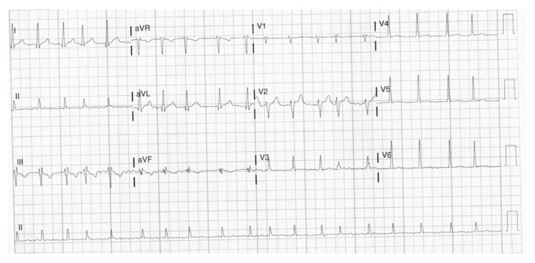
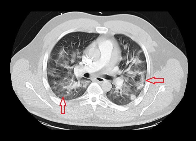
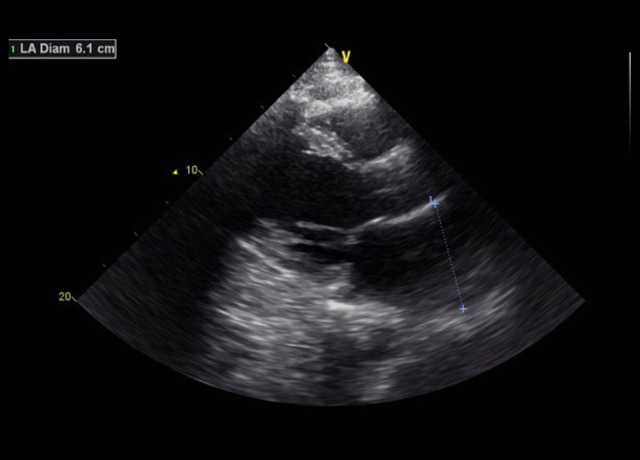
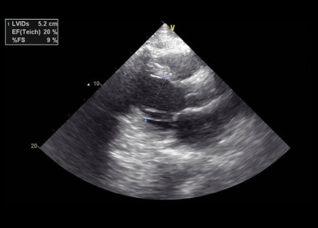
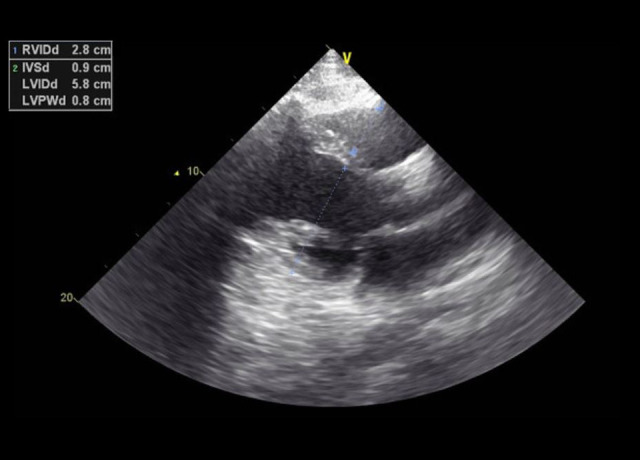
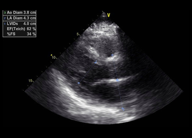
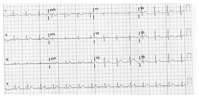
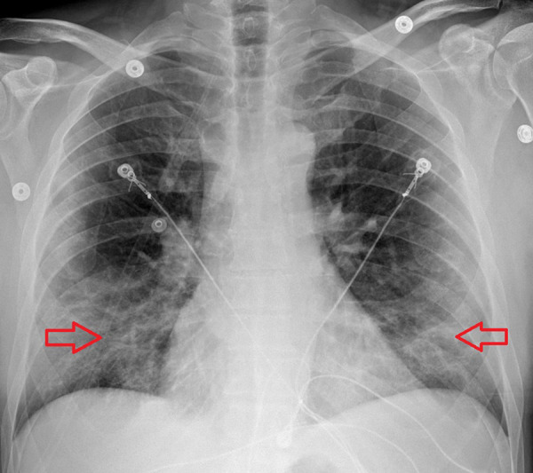
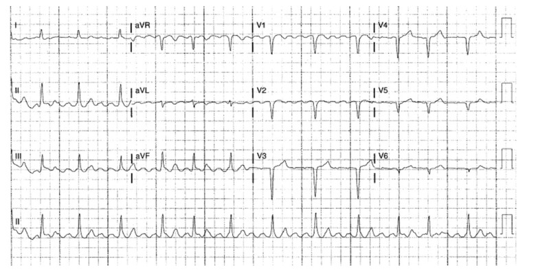
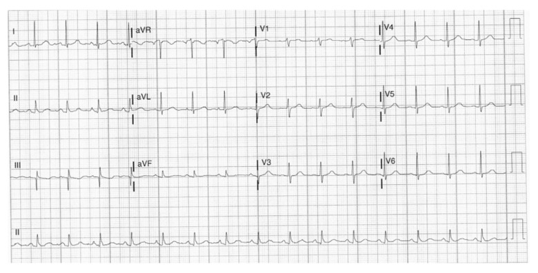
BACKGROUND We report 4 family members, a 29-year-old son, 54-year-old father, 79-year-old grandmother, and 84-year-old grandfather, with COVID-19 pneumonia. Only the son had heart failure, with reduced ejection fraction and atrial fibrillation. This report aims to show that age and baseline comorbidities are not always predictors of severe COVID-19 disease. CASE REPORT Case 1: The son, a 29-year-old man, presented with dyspnea and palpitation. His nasopharyngeal swab was positive for severe acute respiratory syndrome coronavirus 2 (SARS-CoV-2). He required high-flow nasal cannula oxygen therapy and had new-onset atrial fibrillation and reduced ejection fraction. Case 2: The father, a 54-year-old man, presented with dyspnea. Nasopharyngeal swab was positive for SARS-CoV-2. He required regular nasal cannula oxygen therapy. Electrocardiogram showed sinus rhythm. Echocardiogram showed normal ejection fraction. Case 3: The grandfather, an 84-year-old man with a history of atrial flutter, chronic kidney disease, and hypertension, presented with dyspnea and fever. Nasopharyngeal swab was positive for SARS-CoV-2. He required regular nasal cannula oxygen therapy. Electrocardiogram showed sinus rhythm. Echocardiogram showed normal ejection fraction. Case 4: The grandmother, a 79-year-old woman with a history of hypertension, presented with dyspnea. Nasopharyngeal swab was positive for SARS-CoV-2. She required regular nasal cannula oxygen therapy. Electrocardiogram showed sinus rhythm. CONCLUSIONS COVID-19 caused by SARS-CoV-2 is recognized to affect family members and can involve the heart, causing heart failure and cardiac arrhythmia like atrial fibrillation. This report highlights the importance of cardiac monitoring and consideration of cardiac damage, even without previous risk factors, in all hospitalized patients with COVID-19.
Conflict of interest statement
None.
Figures
Similar articles
-
COVID-19-associated myocarditis presenting as new-onset heart failure and atrial fibrillation.BMJ Case Rep. 2021 Jul 12;14(7):e244027. doi: 10.1136/bcr-2021-244027. BMJ Case Rep. 2021. PMID: 34253533 Free PMC article.
-
A rare presentation of a patient with COVID-19: Cardiac tamponade.Turk Kardiyol Dern Ars. 2020 Oct;48(7):703-706. doi: 10.5543/tkda.2020.56727. Turk Kardiyol Dern Ars. 2020. PMID: 33034578 English.
-
Cardiac Involvement in a Patient With Coronavirus Disease 2019 (COVID-19).JAMA Cardiol. 2020 Jul 1;5(7):819-824. doi: 10.1001/jamacardio.2020.1096. JAMA Cardiol. 2020. PMID: 32219357 Free PMC article.
-
A Periaortitis Patient Who Succumbed to COVID-19 While Undergoing Systemic Steroid Therapy: A Case Report and Literature Review.Am J Case Rep. 2021 Aug 20;22:e932733. doi: 10.12659/AJCR.932733. Am J Case Rep. 2021. PMID: 34415896 Free PMC article. Review.
-
Has COVID-19 changed the spectrum of arrhythmias and the incidence of sudden cardiac death?Herz. 2023 Jun;48(3):212-217. doi: 10.1007/s00059-023-05186-2. Epub 2023 Jun 5. Herz. 2023. PMID: 37277617 Free PMC article. Review.
Cited by
-
A Systematic Review of Case Reports of New-Onset Atrial Fibrillation in COVID-19 Patients.Cureus. 2025 Feb 13;17(2):e78938. doi: 10.7759/cureus.78938. eCollection 2025 Feb. Cureus. 2025. PMID: 40091918 Free PMC article. Review.
-
COVID-19 HEART unveiling as atrial fibrillation: pathophysiology, management and future directions for research.Egypt Heart J. 2023 Apr 30;75(1):36. doi: 10.1186/s43044-023-00359-0. Egypt Heart J. 2023. PMID: 37120772 Free PMC article. Review.
References
-
- Johns Hopkins University Medical Center . Global and US Daily COVID-19 Data. 2021. Coronavirus Resource Center. https://coronavirus.jhu.edu.
Publication types
MeSH terms
LinkOut - more resources
Full Text Sources
Medical
Miscellaneous
