. 2021 Jul 22;12(1):4452.
doi: 10.1038/s41467-021-24736-y.

S-nitrosylation-mediated coupling of G-protein alpha-2 with CXCR5 induces Hippo/YAP-dependent diabetes-accelerated atherosclerosis

Affiliations

S-nitrosylation-mediated coupling of G-protein alpha-2 with CXCR5 induces Hippo/YAP-dependent diabetes-accelerated atherosclerosis

Meng-Lin Chao et al. Nat Commun. .

Abstract

Atherosclerosis-associated cardiovascular disease is one of the main causes of death and disability among patients with diabetes mellitus. However, little is known about the impact of S-nitrosylation in diabetes-accelerated atherosclerosis. Here, we show increased levels of S-nitrosylation of guanine nucleotide-binding protein G(i) subunit alpha-2 (SNO-GNAI2) at Cysteine 66 in coronary artery samples from diabetic patients with atherosclerosis, consistently with results from mice. Mechanistically, SNO-GNAI2 acted by coupling with CXCR5 to dephosphorylate the Hippo pathway kinase LATS1, thereby leading to nuclear translocation of YAP and promoting an inflammatory response in endothelial cells. Furthermore, Cys-mutant GNAI2 refractory to S-nitrosylation abrogated GNAI2-CXCR5 coupling, alleviated atherosclerosis in diabetic mice, restored Hippo activity, and reduced endothelial inflammation. In addition, we showed that melatonin treatment restored endothelial function and protected against diabetes-accelerated atherosclerosis by preventing GNAI2 S-nitrosylation. In conclusion, SNO-GNAI2 drives diabetes-accelerated atherosclerosis by coupling with CXCR5 and activating YAP-dependent endothelial inflammation, and reducing SNO-GNAI2 is an efficient strategy for alleviating diabetes-accelerated atherosclerosis.


Conflict of interest statement

The authors declare no competing interests.

Figures

Fig. 1
Fig. 1. S-nitrosylation of GNAI2 is significantly higher in diabetes-accelerated atherosclerosis in vitro and in vivo.
a A biotin-switch assay detected protein S-nitrosylation in HUVECs treated with Mannitol+nLDL or HG (25 mmol/L) and oxLDL (50 μg/mL). One independent experiment was performed. b S-nitrosylation of GNAI2 is significantly higher in coronary arteries of diabetic patients with CAD. The level of SNO-GNAI2 was quantified with total-GNAI2 and normalized to Control patients. n = 5 distinct samples for each group. c S-nitrosylation of GNAI2 is increased in the aortas of LDLr−/− mice treated with STZ and HFD. The level of SNO-GNAI2 was quantified with total-GNAI2 and normalized to Vehicle + NC mice. n = 6 distinct samples for each group. d S-nitrosylation of GNAI2 is markedly increased in HUVECs treated with HG and oxLDL for 24 h. The level of SNO-GNAI2 was quantified with total-GNAI2 and normalized to Mannitol+nLDL-treated cells. n = 3 distinct samples for each group. N.D represents no detected. e S-nitrosylation of GNAI2 is significantly increased in HAECs treated with HG and oxLDL for 24 h. The level of SNO-GNAI2 was quantified with total-GNAI2 and normalized to Mannitol+nLDL-treated HAECs. n = 3 distinct samples for each group. N.D represents no detected. Data are represented as the Mean ± SEM. Unpaired two-tailed Student’s t-test was used for statistical analysis. Source data are provided as a Source Data file.
Fig. 2
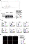
Fig. 2. Inhibition of GNAI2 S-nitrosylation at Cys66 alleviates HG- and oxLDL-induced inflammatory response.
a Cys66 is identified as the SNO-site of GNAI2 according to the liquid chromatography-tandem mass spectrometry (LC-MS/MS) analysis. b HUVECs were ectopically expressed with GNAI2-WT and GNAI2-C66A, followed by exposure to HG and oxLDL for 24 h. GNAI2-C66A abolishes S-nitrosylation of GNAI2 induced by HG and oxLDL as determined by a biotin-switch assay. n = 3 distinct samples for each group. c HAECs were transfected with pcDNA, GNAI2-WT, and GNAI2-C66A, followed by stimulated with HG and oxLDL for 24 h. GNAI2-C66A inhibits the mRNA expressions of adhesion molecules (ICAM1, VCAM1, SELE, and SELP) and chemokines (CXCL4, CXCL8, CCL2, and CCL5) induced by HG and oxLDL as determined by qPCR. n = 3 distinct samples for each group. d GNAI2-C66A prevents the attachment of THP-1 cells to HAECs in HG and oxLDL condition. Scale bar = 100 μm. n = 3 distinct samples for each group. Data are represented as the Mean ± SEM. One-way ANOVA followed by Tukey’s test for post-hoc comparisons was used. Source data are provided as a Source Data file.
Fig. 3
Fig. 3. Inhibition of SNO-GNAI2 at Cys66 mitigates endothelial dysfunction in diabetes-accelerated atherosclerosis.
LDLr−/− mice were transfected with AAVendo-GFP, AAVendo-GNAI2-WT, or AAVendo-GNAI2-C66A, respectively, and randomly separated into NC, HFD, and STZ + HFD groups. a AAVendo-GNAI2-C66A significantly diminishes S-nitrosylation of GNAI2 in the aorta, as determined through a biotin-switch assay. n = 6 mice for each group. N.D represents no detected. b Endothelial specific transfection of GNAI2-C66A alleviates diabetes-accelerated atherosclerosis. Scale bar = 5 mm. n = 6 mice for each group. N.D represents no detected. c Endothelial specific transfection of GNAI2-C66A decreases plaque areas in the aortic roots, evidenced by hematoxylin and eosin staining (H&E). Scale bar = 200 μm. n = 6 mice for each group. N.D represents no detected. d, e AAVendo-GNAI2-C66A increases the stability of atherosclerotic plaques, as evidenced by picrosirius red staining for collagen (Scale bar = 200 μm), α-SMA for smooth muscle cells (Scale bar = 50 μm), CD68 for macrophage infiltration (Scale bar = 50 μm), and Oil Red O staining for lipid deposition (Scale bar = 200 μm), n = 6 mice for each group. N.D represents no detected. f Endothelial specific transfection of GNAI2-C66A rescues the impairment in endothelium-dependent relaxation induced by STZ and HFD in the presence of Ach (10−9 to 10−5 mol/L). n = 6 mice for each group. g The mRNA expressions of adhesion molecules (Icam1, Vcam1, Sele, and Selp) and chemokines (Cxcl1, Cxcl4, Ccl2, and Ccl5) are significantly reduced in aortas of STZ and HFD-treated LDLr−/− mice transfected with AAVendo-GNAI2-C66A. n = 6 mice for each group. Data are represented as the Mean ± SEM. a, b, e, f, g Welch ANOVA followed by Tamhane’s T2 test for post-hoc comparisons. c One-way ANOVA followed by Tukey’s test for post-hoc comparisons. Source data are provided as a Source Data file.
Fig. 4
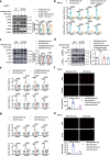
Fig. 4. S-nitrosylation of GNAI2 at Cys66 mediates CXCR5 activation and Hippo-YAP pathway dysfunction.
a Interaction of endogenous GNAI2 with CXCR5. Three indepenent experiments were performed. b GNAI2-C66A markedly decreases the interaction between GNAI2 and CXCR5 induced by HG + oxLDL. n = 3 distinct samples for each group. N.D represents no detected. c, d The BRET assay shows that GNAI2-C66A reverses the reduced EC50 of CXCL13 induced by treatment with DETA. n = 3 distinct samples for each group. e Ectopic expression of GNAI2-C66A rather than GNAI2-WT partly inhibits the reduction of cAMP in HUVECs stimulated with HG + oxLDL. n = 3 independent experiments. f Overexpression of GNAI2-C66A restores the decrease of phospho-LAST1 kinase and phospho-YAP induced by HG and oxLDL. n = 3 distinct samples for each group. g Overexpression of GNAI2-C66A reduces the nuclear level of YAP in HUVECs stimulated with HG + oxLDL. n = 3 distinct samples for each group. h Accumulation of YAP (Red) in nuclei (Blue) is reduced in HUVECs ectopically overexpressed with GNAI2-C66A. HUVECs were stained with CD31 (Green). Scale bar = 20 μm. Three indepenent experiments were performed. i Endothelial specific transduction of GNAI2-C66A rescues the dephosphorylation of LATS1 and YAP induced by STZ and HFD in LDLr−/− mice. n = 6 distinct samples for each group. j Compared to CAD patients, phosphorylated levels of LATS1 and YAP are reduced in coronary arteries of diabetic patients with CAD. n = 5 distinct samples for each group. Data are represented as the Mean ± SEM. b, d, e, f, g, i One-way ANOVA followed by Tukey’s test for post-hoc comparisons. j Unpaired two-tailed Student’s t-test for pLATS1 and Mann–Whitney test for pYAP. Source data are provided as a Source Data file.
Fig. 5
Fig. 5. Inactivation of CXCR5-Hippo-YAP pathway improves endothelial inflammation and monocyte adhesion induced by HG and oxLDL.
a MAECs isolated from CXCR5−/− mice were treated with HG + oxLDL for 24 h. Knockout of CXCR5 reverses the decreased phosphorylation of LATS1 and YAP induced by HG + oxLDL. n = 3 distinct samples for each group. b Knockout of CXCR5 reduces the expression of adhesion molecules and chemokines. n = 3 independent experiments. c HUVECs were transfected with siNC or siCXCR5, subjected to HG and oxLDL for 24 h, the level of phospho-LAST1 and phospho-YAP was determined. Knockdown of CXCR5 restores the phosphorylation of LATS1 and YAP, d reduces YAP nuclear translocation, e suppresses the expression of adhesion molecules and chemokines. c–e n = 5 distinct samples for each group. f Knockdown of CXCR5 reduces the attachment of THP-1 monocytes to HAECs treated with HG and oxLDL. Scale bar = 100 μm. n = 3 distinct samples for each group. g HUVECs were transfected with siNC or siYAP, followed by stimulated with HG and oxLDL. Knockdown of YAP attenuates the mRNA expression of adhesion molecules and inflammatory chemokines. n = 5 distinct samples for each group. h Knockdown of YAP decreases the attachment of THP-1 cells to HG- and oxLDL-stimulated HAECs. n = 3 distinct samples for each group. Data are represented as the Mean ± SEM. VCAM1, CCL2 in e and ICAM1, SELE in g were analyzed by Welch ANOVA followed by Tamhane’s T2 test for post-hoc comparisons. Others were analyzed by One-way ANOVA followed by Tukey’s test for post-hoc comparisons. Source data are provided as a Source Data file.
Fig. 6
Fig. 6. iNOS is responsible for melatonin-induced inhibition of SNO-GNAI2 and melatonin suppresses endothelial inflammation through CXCR5/Hippo pathway.
a HG + oxLDL increases the level of iNOS, rather than eNOS, phospho-eNOS, GSNOR, or Trx. n = 5 cell samples for each group. b The expression of iNOS is increased in coronary arteries of diabetic patients with CAD and in aortas of STZ + HFD-treated LDL−/− mice. n = 7 human samples. n = 5 mice samples. c 1400 W (100 μM) pretreatment for 15 min abolishes the elevation of SNO-GNAI2 induced by HG + oxLDL in HUVECs. n = 3 independent experiments. N.D represents no detected. d 1400 W represses the mRNA expressions of adhesion molecules and chemokines. n = 3 independent experiments. e Melatonin dose-dependently (1 μM, 2 μM, 5 μM, 10 μM, 20 μM) reduces the level of iNOS in HG + oxLDL-treated HUVECs. n = 3 independent experiments. f Melatonin decreases S-nitrosylation of GNAI2 in HG + oxLDL-treated HUVECs. n = 5 independent experiments. N.D represents no detected. g Melatonin represses the interaction between GNAI2 and CXCR5 (n = 3 independent experiments, N.D represents no detected), h reverses the dephosphorylation of LATS1 and YAP (n = 4 independent experiments) and i reduces the nuclear translocation of YAP in HUVECs treated with HG + oxLDL (n = 3 independent experiments). j Melatonin reduces nuclear translocation of YAP. YAP (red), nuclei (DAPI, blue), and HUVECs (CD31, green). Scale bar = 20 μm. Three indepenent experiments were performed. k Melatonin abolishes the elevation of adhesion molecules and chemokines in HAECs induced by HG + oxLDL. l Melatonin attenuates the adhesion of THP-1 cells to HAECs. Scale bar = 100 μm. k, l n = 3 independent experiments. Data are represented as the Mean ± SEM. a Mann–Whitney test. b Mann–Whitney test for mice samples and Unpaired two-tailed Student’s t-test for human samples. c, d, f, g, h, i, k, l One-way ANOVA followed by Tukey’s test for post-hoc comparisons. e One-way ANOVA followed by Dunnett’s test for post-hoc comparisons. Source data are provided as a Source Data file.
Fig. 7
Fig. 7. Melatonin exerts anti-inflammation and athero-protective role under diabetic condition.
a Melatonin decreases the S-nitrosylation of GNAI2 in aortas of STZ + HFD-treated LDLr−/− mice. n = 6 mice for each group. b Melatonin mitigates the diabetes-accelerated atherosclerosis as evidenced by en face Oil Red O staining of the whole aortas and melatonin mainly reduces the lesion in aortic arch. Scale bar = 5 mm. n = 6 mice for each group. N.D represents no detected. c Melatonin attenuates the plaque areas in aortic roots induced by STZ + HFD. scale bar = 200 μm. n = 7 mice in STZ + HFD + MLT group and n = 6 mice in other groups. N.D represents no detected. d Melatonin increases collagen deposition and smooth muscle cell content, reduces macrophage infiltration and lipid accumulation in aortas of STZ and HFD-treated LDLr−/− mice, as determined by PSR staining (scale bar = 200 μm), immunofluorescent staining of α-SMA and CD68+ macrophage (scale bar = 50 μm), and Oil Red O staining (scale bar =200 μm). e Quantification of collagen content (n = 7 mice in STZ + HFD + MLT group and n = 6 mice in other groups), smooth muscle cell content (n = 6 mice for each group), macrophage infiltration (n = 6 mice for each group), and lipid accumulation (n = 6 mice for each group). N.D represents no detected. f Melatonin rescues the impairment in Ach-mediated endothelium-dependent relaxation in STZ + HFD-treated LDLr−/− mice. n = 6 mice for each group. g Melatonin suppresses the mRNA levels of adhesion molecules and chemokines in STZ + HFD-treated LDLr−/− mice. n = 6 mice for each group. h Melatonin restores the phosphorylation of LATS1 and YAP in the aortas of STZ + HFD-treated LDLr−/− mice. n = 6 mice for each group. Data are represented as the Mean ± SEM. a, b, e, g, h Welch ANOVA followed by Tamhane’s T2 test for post-hoc comparisons. For Icam1 in g, and c, f One-way ANOVA followed by Turkey’s test for post-hoc comparisons was used. Source data are provided as a Source Data file.
Fig. 8
Fig. 8. Graphic model of SNO-GNAI2-induced diabetes-accelerated atherosclerosis.
During diabetes-accelerated atherosclerosis, high glucose, and oxLDL increases S-nitrosylation of GNAI2 in endothelial cells, which enhances coupling with CXCR5 and reduces cAMP level. The reduction in cAMP dephosphorylates LATS1 and YAP, promotes nuclear translocation of YAP and promotes transcription of adhesion molecules and chemokines, which enhances endothelial inflammation. Melatonin restores phosphorylation of LAST1 and YAP through reducing iNOS-induced SNO-GNAI2 and alleviates endothelial inflammation, which improves diabetes-accelerated atherosclerosis. oxLDL oxidized low-density lipoprotein, GNAI2 guanine nucleotide-binding protein G(i) subunit alpha-2, CXCR5 C-X-C chemokine receptor type 5, cAMP cyclic adenosine monophosphate, LATS1 large tumor suppressor kinase 1, YAP Yes-associated protein, iNOS inducible nitric oxide synthase.

References

    1. Low Wang CC, Hess CN, Hiatt WR, Goldfine AB. Clinical update: cardiovascular disease in Diabetes mellitus: atherosclerotic cardiovascular disease and heart failure in type 2 Diabetes mellitus—mechanisms, management, and clinical considerations. Circulation. 2016;133:2459–2502. doi: 10.1161/CIRCULATIONAHA.116.022194. - DOI - PMC - PubMed
    1. Funk, S. D., Yurdagul, A., Jr. & Orr, A. W. Hyperglycemia and endothelial dysfunction in atherosclerosis: lessons from type 1 diabetes. Int. J. Vasc. Med. 2012, 569654 (2012). - PMC - PubMed
    1. Kannel WB, McGee DL. Diabetes and cardiovascular disease. The Framingham study. JAMA. 1979;241:2035–2038. doi: 10.1001/jama.1979.03290450033020. - DOI - PubMed
    1. Renard CB, et al. Diabetes and diabetes-associated lipid abnormalities have distinct effects on initiation and progression of atherosclerotic lesions. J. Clin. Invest. 2004;114:659–668. doi: 10.1172/JCI200417867. - DOI - PMC - PubMed
    1. Dahl-Jorgensen K, Larsen JR, Hanssen KF. Atherosclerosis in childhood and adolescent type 1 diabetes: early disease, early treatment? Diabetologia. 2005;48:1445–1453. doi: 10.1007/s00125-005-1832-1. - DOI - PubMed

Publication types

MeSH terms