

Clinically applicable artificial intelligence system for dental diagnosis with CBCT
- PMID: 34294759
- PMCID: PMC8298426
- DOI: 10.1038/s41598-021-94093-9
Clinically applicable artificial intelligence system for dental diagnosis with CBCT
Erratum in
-
Author Correction: Clinically applicable artificial intelligence system for dental diagnosis with CBCT.Sci Rep. 2021 Nov 9;11(1):22217. doi: 10.1038/s41598-021-01678-5. Sci Rep. 2021. PMID: 34754062 Free PMC article. No abstract available.
Abstract
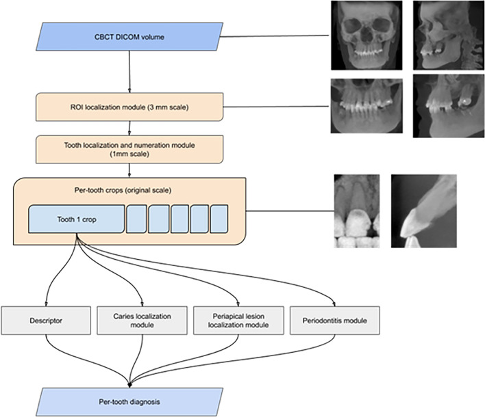
In this study, a novel AI system based on deep learning methods was evaluated to determine its real-time performance of CBCT imaging diagnosis of anatomical landmarks, pathologies, clinical effectiveness, and safety when used by dentists in a clinical setting. The system consists of 5 modules: ROI-localization-module (segmentation of teeth and jaws), tooth-localization and numeration-module, periodontitis-module, caries-localization-module, and periapical-lesion-localization-module. These modules use CNN based on state-of-the-art architectures. In total, 1346 CBCT scans were used to train the modules. After annotation and model development, the AI system was tested for diagnostic capabilities of the Diagnocat AI system. 24 dentists participated in the clinical evaluation of the system. 30 CBCT scans were examined by two groups of dentists, where one group was aided by Diagnocat and the other was unaided. The results for the overall sensitivity and specificity for aided and unaided groups were calculated as an aggregate of all conditions. The sensitivity values for aided and unaided groups were 0.8537 and 0.7672 while specificity was 0.9672 and 0.9616 respectively. There was a statistically significant difference between the groups (p = 0.032). This study showed that the proposed AI system significantly improved the diagnostic capabilities of dentists.
© 2021. The Author(s).
Conflict of interest statement
Financial support was received by Diagnocat Co. Ltd., San Francisco CA. Matvey Ezhov, Maxim Gusarev, Maria Golitsyna, Eugene Shumilov, and Alex Sanders are employees of Diagnocat Co. Ltd. Kaan Orhan is a scientific research advisor for the Diagnocat Co. Ltd., San Francisco CA. Julian M Yates, Evgeny Kushnerev, Dania Tamimi, Secil Aksoy have no potential competing interests.
Figures


References
-
- White SC, Pharoah MJ. Oral Radiology-E-Book: Principles and Interpretation. Elsevier Health Sciences; 2014.
-
- Oz U, Orhan K, Abe N. Comparison of linear and angular measurements using two-dimensional conventional methods and three-dimensional cone beam CT images reconstructed from a volumetric rendering program in vivo. Dentomaxillofac. Radiol. 2011;40(8):492–500. doi: 10.1259/dmfr/15644321. - DOI - PMC - PubMed
Publication types
MeSH terms
LinkOut - more resources
Full Text Sources
