. 2021 Jul 22;11(1):15047.
doi: 10.1038/s41598-021-94377-0.

Autoantibody profiles and clinical association in Thai patients with autoimmune retinopathy

Affiliations

Autoantibody profiles and clinical association in Thai patients with autoimmune retinopathy

Aulia Rahmi Pawestri et al. Sci Rep. .

Abstract

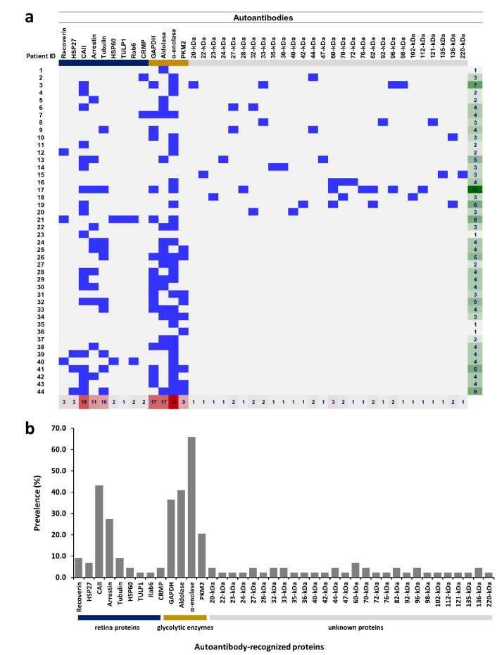
Autoimmune retinopathy (AIR) is a rare immune-mediated inflammation of the retina. The autoantibodies against retinal proteins and glycolytic enzymes were reported to be involved in the pathogenesis. This retrospective cohort study assessed the antiretinal autoantibody profiles and their association with clinical outcomes of AIR patients in Thailand. We included 44 patients, 75% were females, with the overall median age of onset of 48 (17-74, IQR 40-55.5) years. Common clinical presentations were nyctalopia (65.9%), blurred vision (52.3%), constricted visual field (43.2%), and nonrecordable electroretinography (65.9%). Underlying malignancy and autoimmune diseases were found in 2 and 12 female patients, respectively. We found 41 autoantibodies, with anti-α-enolase (65.9%) showing the highest prevalence, followed by anti-CAII (43.2%), anti-aldolase (40.9%), and anti-GAPDH (36.4%). Anti-aldolase was associated with male gender (P = 0.012, OR 7.11, 95% CI 1.54-32.91). Anti-CAII showed significant association with age of onset (P = 0.025, 95% CI - 17.28 to - 1.24), while anti-α-enolase (P = 0.002, OR 4.37, 95% CI 1.83-10.37) and anti-GAPDH (P = 0.001, OR 1.87, 95% CI 1.32-2.64) were significantly associated with nonrecordable electroretinography. Association between the antibody profiles and clinical outcomes may be used to direct and adjust the treatment plans and provide insights in the pathogenesis of AIR.


Conflict of interest statement

The authors declare no competing interests.

Figures

Figure 1
Figure 1
Disease progression of a patient with autoimmune retinopathy (AIR). Color fundus photography at baseline in the right (a) and left eye (b) shows normal optic disc, arteriolar attenuation, generalized retinal pigment epithelium (RPE) atrophy with macular sparing and scattered pigment clumps. Prominent large choroidal vessels can be observed around the optic disc. Optical coherence tomography images at baseline showing RPE attenuation, loss of ellipsoid zone (EZ) at the periphery, flattening of the outer nuclear layer (ONL), and slit cavitation in the inner nuclear layer (INL) on the nasal side, as marked by arrows, in the right (c) and left eye (d). The disease progression after five years is marked with RPE atrophy, progressive loss of EZ and ONL toward the fovea, more prominent slit cavitation in the INL, as marked by arrows, and accompanying choroidal thinning in the right (e) and left eye (f). The data is retrieved from patient number 1.
Figure 2
Figure 2
(a) Autoantibody profile in patients. The antibody array depicts the positive autoantibodies against the corresponding proteins (blue boxes), negative results are shown as grey boxes and white boxes indicate that the test was not assigned. The colored line under the antibody names described the retina proteins (dark blue), glycolytic enzymes (yellow), and unidentified retina proteins (grey). The last column on the right describes the total number of antibodies in each patient (green gradient) and the lowest row shows the number of patients having each type of antibody (red gradient). (b) Prevalence of autoantibodies in 44 patients. Bar chart depicts the prevalence of the autoantibodies based on the three protein panels: retina proteins (dark blue), glycolytic enzyme (yellow), and unidentified retina proteins (grey). HSP heat shock protein, CAII carbonic anhydrase II, TULP1 tubby-like protein 1, CRMP collapsin response mediator protein, GAPDH glyceraldehyde 3-phosphate dehydrogenase, PKM2 pyruvate kinase isozyme M2, kDa kilo Dalton.

References

    1. Adamus G. Are anti-retinal autoantibodies a cause or a consequence of retinal degeneration in autoimmune retinopathies? Front. Immunol. 2018;9:765. doi: 10.3389/fimmu.2018.00765. - DOI - PMC - PubMed
    1. Adamus G, Champaigne R, Yang S. Occurrence of major anti-retinal autoantibodies associated with paraneoplastic autoimmune retinopathy. Clin. Immunol. 2020;210:108317. doi: 10.1016/j.clim.2019.108317. - DOI - PMC - PubMed
    1. Braithwaite T, Vugler A, Tufail A. Autoimmune retinopathy. Ophthalmologica. 2012;228:131–142. doi: 10.1159/000338240. - DOI - PubMed
    1. Sen HN, Grange L, Akanda M, Fox A. Autoimmune retinopathy: Current concepts and practices (An American Ophthalmological Society thesis) Trans. Am. Ophthalmol. Soc. 2017;115:T8. - PMC - PubMed
    1. Heckenlively JR, Ferreyra HA. Autoimmune retinopathy: A review and summary. Semin. Immunopathol. 2008;30:127–134. doi: 10.1007/s00281-008-0114-7. - DOI - PubMed

Publication types

MeSH terms