. 2022 Jan;29(1):82-95.
doi: 10.1038/s41418-021-00838-4. Epub 2021 Jul 22.

Exosomal miR-101-3p and miR-423-5p inhibit medulloblastoma tumorigenesis through targeting FOXP4 and EZH2

Affiliations

Exosomal miR-101-3p and miR-423-5p inhibit medulloblastoma tumorigenesis through targeting FOXP4 and EZH2

Ping Xue et al. Cell Death Differ. 2022 Jan.

Erratum in

Abstract

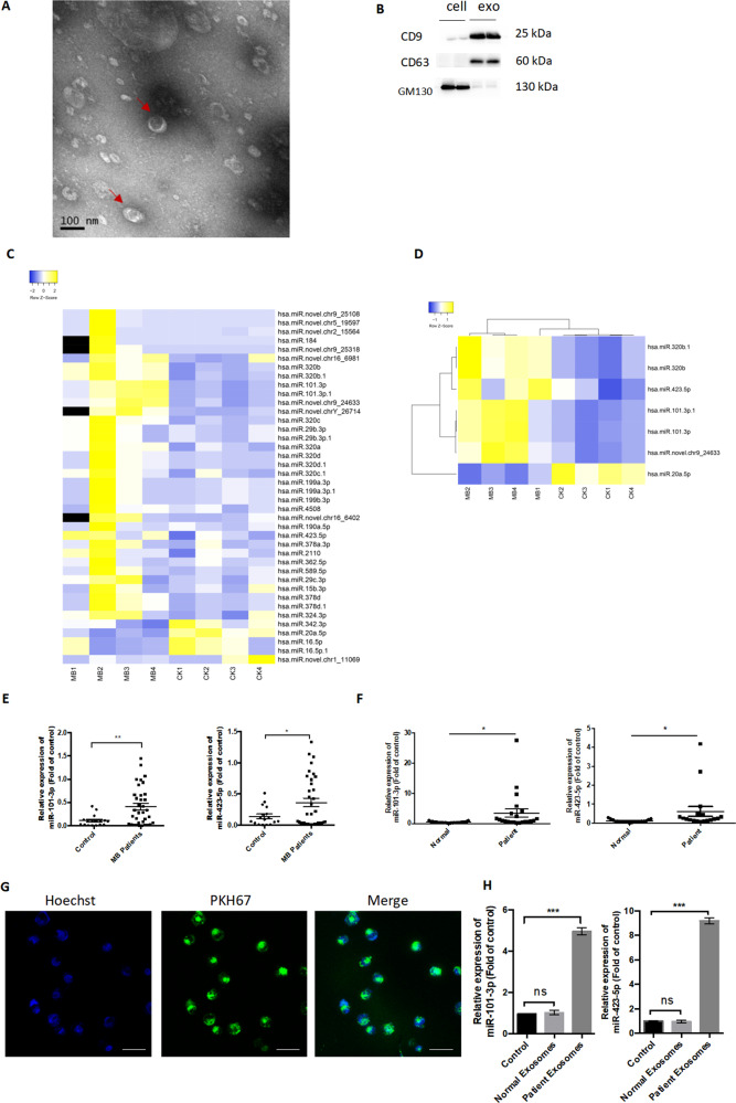
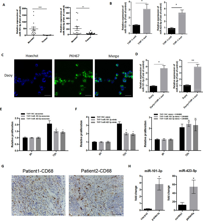
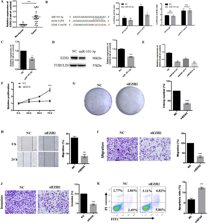
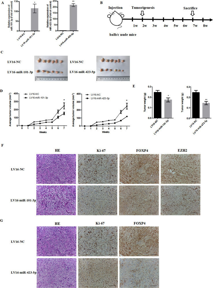
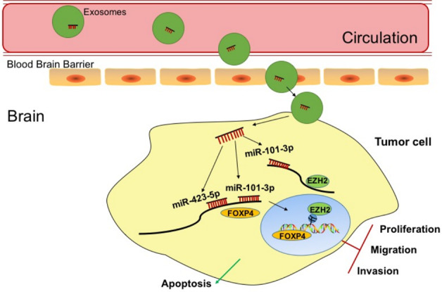
Exosomal microRNAs (miRNAs) have been implicated in the development and progression of a variety of tumors; however, whether they contribute to medulloblastoma (MB) tumorigenesis remains to be elucidated. To address this, we first characterized the miRNA profiles of circulating exosomes by miRNA sequencing to identify miRNAs differentially expressed between children with MB and healthy controls. Then, we conducted in vitro and in vivo functional assays with the identified miRNAs and their predicted targets. We found that, compared with healthy controls, 35 miRNAs were upregulated and 5 downregulated in exosomes isolated from the plasma of MB patients. We further found that the expression of miR-101-3p and miR-423-5p was significantly higher in plasma exosomes from MB patients than in healthy controls in an expanded cohort and these exosomal miRNAs could be delivered to tumor cells via exosomes. An in vitro functional analysis of miR-101-3p and miR-423-5p showed that treating MB cells with the corresponding mimics significantly inhibited the proliferation, colony-forming ability, migratory ability, and invasive capacity of tumor cells, and promoted cell apoptosis. Additionally, miR-101-3p and miR-423-5p were found to act as tumor suppressors by directly targeting a common gene, FOXP4, which encodes a transcription factor with a vital role in embryonic development and tumorigenesis. Moreover, miR-101-3p also targeted EZH2, a histone methyltransferase, to reinforce its tumor inhibitory effects. Using a xenograft nude mouse model of MB, we further identified that the overexpression of miR-101-3p and miR-423-5p inhibited tumorigenesis in vivo. Our findings provide novel insights into the functions of exosomal miRNAs in mediating MB progression and suggest a potential therapeutic approach for the treatment of children with MB.


Conflict of interest statement

The authors declare no competing interests.

Figures

Fig. 1
Fig. 1. Profiles of differentially expressed exosomal miRNAs in patients with medulloblastoma.
A Transmission electron microscopy analysis. Scale bar, 100 nm. B Western blotting analysis of the exosomal marker proteins CD9, CD63, and GM130. C Heatmap showing exosomal miRNAs differentially expressed between medulloblastoma (MB) patients and healthy controls (fold change >1.5). D Heatmap showing exosomal miRNAs differentially expressed at fold change >1.5 and p < 0.05. E, F RT-qPCR-based verification of miR-101-3p and miR-423-5p expression levels in plasma-derived exosomes (E) or peripheral blood mononuclear cells (PBMCs) (F) in an expanded cohort (MB = 35, control = 17). G Internalization of plasma-derived exosomes into Daoy cells. Scale bar indicates 50 µm. H The expression levels of miR-101-3p and miR-423-5p in Daoy cells after culture with plasma-derived exosomes isolated from MB patients and normal controls. Data are presented as means ± SEM. *p < 0.05, **p < 0.01, ***p < 0.001.
Fig. 2
Fig. 2. The overexpression of miR-101-3p and miR-423-5p inhibited cell proliferation, migration, and invasion, and promoted cell apoptosis.
A, B Cell Counting Kit-8 (CCK-8) assays showing that Daoy and D283 Med cells transfected with miR-101-3p/miR-423-5p mimics or lentivirus exhibit decreased cell proliferation compared with cells transfected with miR-NC (negative control). C A CCK-8 cell proliferation assay was performed on Daoy and D283 Med cells cultured with pooled exosomes (50 µg/ml) derived from the plasma of medulloblastoma patients. D, E Flow cytometric analysis of cell apoptosis in Daoy and D283 Med cells transfected with miR-101-3p or miR-423-5p mimics or lentiviral vectors. F MiR-101-3p and miR-423-5p mimics inhibited Daoy cell migration into wound areas. Scale bar indicates 100 µm. G, H Transwell assays showing that the migratory and invasive abilities of Daoy cells transfected with miR-101-3p or miR-423-5p mimics are inhibited compared with those of cells transfected with miR-NC. Scale bar indicates 100 µm. Data are presented as means ± SEM. *p < 0.05, **p < 0.01, ***p < 0.001.
Fig. 3
Fig. 3. Monocyte/macrophage cell-derived exosomal miR-101-3p and miR-423-5p could be transferred into medulloblastoma cells.
A The expression levels of miR-101-3p and miR-423-5p in tumor tissues from medulloblastoma (MB) patients relative to those in adjacent normal brain tissues (MB = 25, control = 15). B The expression of miR-101-3p and miR-423-5p in THP-1 cells and exosomes derived from THP-1 cell culture supernatants. C Exosomes from THP-1 cell culture supernatants cultured with Daoy cells. Scale bar indicates 50 µm. D The expression of miR-101-3p and miR-423-5p in Daoy cells cultured with THP-1-derived exosomes. E The proliferative ability of Daoy cells cultured with exosomes (50 µg/ml) derived from supernatant of THP-1 cells transfected with miR-101-3p/miR-423-5p or miR-NC (negative control). F The proliferative ability of Daoy cells cultured with supernatant of THP-1 cells transfected with miR-101-3p/miR-423-5p mimics or miR-NC (negative control) and treated or not with GW4869 (40 µM). G Immunohistochemical analysis of CD68 expression in MB tissue. Scale bar indicates 100 µm. H Compared with that in the controls, the expression of exosomal miR-101-3p and miR-423-5p was higher in MB patient-derived CD14+ monocyte culture supernatant. Data are presented as means ± SEM. *p < 0.05, **p < 0.01, ***p < 0.001.
Fig. 4
Fig. 4. FOXP4 is a target of both miR-101-3p and miR-423-5p.
A A Venn diagram showing target genes shared between miR-101-3p and miR-423-5p, as predicted from two databases (TargetScan and RNA22). B Western blot analysis of FOXP4 protein expression in Daoy cells transfected with the miR-101-3p mimic or miR-NC (negative control). C Western blot analysis of FOXP4 protein expression in Daoy cells transfected with the miR-423-5p mimic or miR-NC. D The predicted binding sites of miR-101-3p and miR-423-5p in the FOXP4- wild or mutated 3′UTR (left). Dual-luciferase assay in HEK293T cells co-transfected with the miR-101-3p or miR-423-5p mimic and a construct containing the wild-type or mutated FOXP4-3′UTR (right). Data are presented as means ± SEM. *p < 0.05, **p < 0.01, ***p < 0.001.
Fig. 5
Fig. 5. FOXP4 functions as an oncogene in medulloblastoma cells.
A Comparison of the expression levels of FOXP4 in tumor samples from medulloblastoma (MB) patients and those in adjacent normal tissues as determined by qPCR (MB = 25, control = 15). B The knockdown of FOXP4 in Daoy cells using siRNAs; siFOXP4-1001 was selected for use in subsequent experiments. C, D The knockdown of FOXP4 inhibited cell proliferation and clonogenicity in Daoy cells. E Wound-healing assay showing that knockdown of FOXP4 using siFOXP4-1001 inhibited cell migration into wound areas. Scale bar indicates 100 µm. F, G Transwell assays showing that the knockdown of FOXP4 decreased cell migration and invasion Scale bar indicates 100 µm. H The knockdown of FOXP4 promoted cell apoptosis, as determined by flow cytometry. Data are presented as means ± SEM. *p < 0.05, **p < 0.01, ***p < 0.001.
Fig. 6
Fig. 6. EZH2 is a direct target of miR-101-3p and functions as an oncogene in medulloblastoma cells.
A Relative EZH2 mRNA expression in tumor samples (n = 25) compared with that in normal tissues (n = 15). B Predicted binding site of miR-101-3p in the EZH2- wild or mutated 3′UTR (left). Luciferase reporter assay in HEK293T cells co-transfected with the miR-101-3p mimic and a construct containing the wild-type or mutated EZH2 3′UTR (right). C EZH2 mRNA expression in Daoy cells transfected with the miR-101-3p mimic. D EZH2 protein level in Daoy cells transfected with the miR-101-3p mimic. E Knockdown of EZH2 by siRNAs; siEZH2-614 was selected for use in subsequent experiments. F–G Cell proliferation and clonogenicity were significantly inhibited in Daoy cells transfected with siEZH2. H The knockdown of EZH2 significantly inhibited cell migration into wound areas in Daoy cells. Scale bar indicates 100 µm. I, J Transwell assays showing that migratory and invasive abilities are inhibited in Daoy cells transfected with siEZH2. Scale bar indicates 100 µm. K Flow cytometry results showing that cell apoptosis rates were increased in Daoy cells transfected with siEZH2. Data are presented as means ± SEM from three independent experiments. *p < 0.05, **p < 0.01, ***p < 0.001.
Fig. 7
Fig. 7. The overexpression of miR-101-3p and miR-423-5p inhibited tumorigenesis in vivo.
A Relative expression levels of miR-101-3p and miR-423-5p in Daoy cells transfected with LV16-miR-101-3p or LV16-miR-423-5p compared with those in cells transfected with LV16-NC (negative control). B Daoy cells transfected with LV16-miR-101-3p, LV16-miR-423-5p, or LV16-NC were subcutaneously injected into Balb/c nude mice and tumor volumes were measured weekly (n = 6/group/experiment). C Images of tumors (LV16-miR-101-3p/LV16-miR-423-5p vs. LV16-NC). D Growth curves of tumor volumes in the different groups of mice. E Tumor weights of mice at week 7 postinjection. F, G Hematoxylin and eosin staining of tumors and the expression of Ki-67, FOXP4, and EZH2 as determined by immunohistochemistry. Scale bar indicates 100 µm. The animal experiments were performed in duplicate. Data are presented as means ± SEM. *p < 0.05, **p < 0.01.
Fig. 8
Fig. 8. MiR-101-3p and miR-423-5p inhibit medulloblastoma tumorigenesis through the downregulation of FOXP4 and EZH2.
Diagram of the functions of miR-101-3p and miR-423-5p in medulloblastoma.

References

    1. Kohler BA, Ward E, McCarthy BJ, Schymura MJ, Ries LA, Eheman C, et al. Annual report to the nation on the status of cancer, 1975-2007, featuring tumors of the brain and other nervous system. J Natl Cancer Inst. 2011;103:714–36. 10.1093/jnci/djr077 - DOI - PMC - PubMed
    1. Massimino M, Biassoni V, Gandola L, Garre ML, Gatta G, Giangaspero F. et al. Childhood medulloblastoma. Crit Rev Oncol Hematol. 2016;105:35–51. 10.1016/j.critrevonc.2016.05.012 - DOI - PubMed
    1. Vlassov AV, Magdaleno S, Setterquist R, Conrad R. Exosomes: current knowledge of their composition, biological functions, and diagnostic and therapeutic potentials. Biochim. Biophys. Acta Gen. Subj. 2012;1820:940–8.10.1016/j.bbagen.2012.03.017 - DOI - PubMed
    1. Mathivanan S, Fahner CJ, Reid GE, Simpson RJ. ExoCarta 2012: database of exosomal proteins, RNA and lipids. Nucleic Acids Res. 2012;40:D1241–4. 10.1093/nar/gkr828 - DOI - PMC - PubMed
    1. Leichter AL, Sullivan MJ, Eccles MR, Chatterjee A. MicroRNA expression patterns and signalling pathways in the development and progression of childhood solid tumours. Mol Cancer. 2017;16:15. 10.1186/s12943-017-0584-0 - DOI - PMC - PubMed

Publication types