Review
. 2021 Jun;113(3):170-184.
doi: 10.32074/1591-951X-245.

Autoimmune biliary diseases: primary biliary cholangitis and primary sclerosing cholangitis

Affiliations
Review

Autoimmune biliary diseases: primary biliary cholangitis and primary sclerosing cholangitis

Samantha Sarcognato et al. Pathologica. 2021 Jun.

Abstract

Autoimmune cholestatic liver diseases are rare hepato-biliary disorders characterized by a progressive, inflammatory destruction of bile ducts. Primary biliary cholangitis (PBC) and primary sclerosing cholangitis (PSC) are the main autoimmune cholestatic liver diseases. Both may evolve into secondary biliary cirrhosis and its complications. Therapeutic options are limited and liver transplantation remains the only definitive treatment for PBC and PSC.

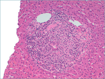
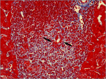
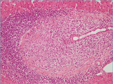
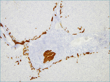
Most PBC and PSC patients have a typical presentation, which does not require liver biopsy. However, in routine clinical practice, important variants or specific subgroups that benefit from liver biopsy for proper management may be observed. Herein, we provide a general overview of clinical and pathological characteristic of PBC and PSC, highlighting the most important features for routine diagnostic practice.

Keywords: PBC; PSC; autoimmune cholangitis; immune-mediated cholangitis; overlap variants.


Conflict of interest statement

Conflict of interest

The Authors declare no conflict of interest.

Figures

Figure 1.
Figure 1.
A florid duct lesion in a primary biliary cholangitis patient. A chronic inflammatory infiltrate made of lymphocytes and plasma cells surrounds and destroys the bile duct (hematoxylin-eosin; original magnification 20x).
Figure 2.
Figure 2.
Lymphocytes infiltrate between adjacent cholangiocytes, destroying the bile duct basement membrane (arrows) (Masson’s trichrome stain; original magnification 40x).
Figure 3.
Figure 3.
A typical epithelioid non-caseating granuloma in a portal tract, intimately associated with a damaged bile duct (hematoxylin-eosin; original magnification 20x).
Figure 4.
Figure 4.
A portal tract showing a damaged bile duct and diffuse ductular reaction (cytokeratin 7 immunostain; original magnification 10x).
Figure 5.
Figure 5.
A portal tract with dense chronic inflammatory infiltrate, with numerous eosinophils (arrows), focally arranged in clusters (asterisk) (hematoxylin-eosin; original magnification 40x).
Figure 6.
Figure 6.
A liver biopsy from a primary biliary cholangitis patient showing diffuse hepatocellular cholestasis, associated with feathery degeneration of hepatocyte cytoplasm (arrow) (hematoxylin-eosin; original magnification 63x).
Figure 7.
Figure 7.
Ductopenia in a patient with advanced primary biliary cholangitis. Cytokeratin 7 stain shows diffuse ductular reaction, with no native bile ducts left (cytokeratin 7 immunostain; original magnification 5x).
Figure 8.
Figure 8.
Cytokeratin 7 stain highlights biliary metaplasia of hepatocytes (cytokeratin 7 immunostain; original magnification 10x).
Figure 9.
Figure 9.
Biliary-type cirrhosis is characterized by parenchymal nodules with edema at the periphery, giving rise to the so-called halo effect (A). Trichrome stain shows complete dense fibrous septa, and better highlights the halo effect at the parenchyma-septum border (B) (A: hematoxylin-eosin; original magnification 5x; B: Masson’s trichrome stain; original magnification 5x).
Figure 10.
Figure 10.
Minimal change primary biliary cholangitis. Cytokeratin 19 immunostaining shows a decreased number of canals of Hering in an otherwise normal portal tract (cytokeratin 19 immunostain; original magnification 20x).
Figure 11.
Figure 11.
Representation of grading of cholangitis and hepatitis activity, according to Nakanuma’s system .
Figure 12.
Figure 12.
Representation of disease staging, according to Nakanuma’s system .
Figure 13.
Figure 13.
Large intrahepatic bile ducts showing obliterative concentric periductal fibrosis, also called onion skin fibrosis, in a liver biopsy from a primary sclerosing cholangitis patient (hematoxylin-eosin; original magnification 20x).
Figure 14.
Figure 14.
A portal tract showing a bile duct scar (arrow) in a liver biopsy from a primary sclerosing cholangitis patient (A). Van Gieson stain confirms that the bile duct has been replaced by fibrous tissue (arrow) (B) (A: hematoxylin-eosin; original magnification 10x; B: Van Gieson stain; original magnification 20x).
Figure 15.
Figure 15.
Periodic acid-Schiff stain with diastase shows a moderate bile duct basement membrane thickening (arrow) in primary sclerosing cholangitis (PAS-diastase stain; original magnification 40x).
Figure 16.
Figure 16.
Enlarged arterioles with thickened wall and arterial fibrointimal hyperplasia are common findings in primary sclerosing cholangitis (hematoxylin-eosin; original magnification 40x).

References

    1. Lazaridis KN, LaRusso NF. The cholangiopathies. Mayo Clin Proc 2015;90:791-800. https://doi.org/10.1016/j.mayocp.2015.03.017 10.1016/j.mayocp.2015.03.017 - DOI - PMC - PubMed
    1. Lazaridis KN, Strazzabosco M, Larusso NF. The cholangiopathies: disorders of biliary epithelia. Gastroenterology 2004;127:1565-1577. https://doi.org/10.1053/j.gastro.2004.08.006 10.1053/j.gastro.2004.08.006 - DOI - PubMed
    1. Dalekos GN, Gatselis NK. Variant and specific forms of autoimmune cholestatic liver diseases. Arch Immunol Ther Exp (Warsz) 2019;67:197-211. https://doi.org/10.1007/s00005-019-00550-9 10.1007/s00005-019-00550-9 - DOI - PubMed
    1. Harada K, Sato Y, Itatsu K, et al. . Innate immune response to double-stranded RNA in biliary epithelial cells is associated with the pathogenesis of biliary atresia. Hepatology 2007;46:1146-1154. https://doi.org/10.1002/hep.21797 10.1002/hep.21797 - DOI - PubMed
    1. Nakanuma Y, Zen Y. Pathology and immunopathology of immunoglobulin G4-related sclerosing cholangitis: the latest addition to the sclerosing cholangitis family. Hepatol Res 2007;37:S478-486. https://doi.org/10.1111/j.1872-034X.2007.00243.x 10.1111/j.1872-034X.2007.00243.x - DOI - PubMed

MeSH terms