Review
. 2021 Jun;113(3):230-251.
doi: 10.32074/1591-951X-309.

Mesenchymal tumours of the gastrointestinal tract

Affiliations
Review

Mesenchymal tumours of the gastrointestinal tract

Marta Sbaraglia et al. Pathologica. 2021 Jun.

Abstract

Mesenchymal tumours represent a heterogenous group of neoplasms encopassing benign, intermediate malignancy, and malignant entities. Sarcomas account for approximately 1% of human malignancies. In consideration of their rarity as well as of intrinsic complexity, diagnostic accuracy represents a major challenge. Traditionally, mesenchymal tumours are regarded as lesions the occurrence of which is mostly limited to somatic soft tissues. However, the occurrence of soft tissue tumours at visceral sites represent a well recognized event, and the GI-tract ranks among the most frequently involved visceral location. There exist entities such as gastrointestinal stromal tumours (GIST) and malignant gastointestinal neuroectodermal tumors that exhibit exquisite tropism for the GI-tract. This review will focus also on other relevant clinico-pathologic entities in which occurrence at visceral location is not at all negligible.

Keywords: GIST; gastrointestinal tract; mesenchymal tumours; rare tumours; sarcoma.


Conflict of interest statement

Conflict of interest

The Authors declare no conflict of interest.

Figures

Figure 1.
Figure 1.
Spindle cell GIST. The neoplasm is composed of short fascicles of uniform spindle cells, with oval nuclei, eosinophilic cytoplasm and poorly circumscribed borders.
Figure 2.
Figure 2.
Epithelioid GIST. The neoplasm shows round, epithelioid cells with oval shaped nuclei and eosinophilic to clear cytoplasm. The growth pattern is usually described as nested.
Figure 3.
Figure 3.
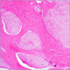
SDH-deficient GIST. This category of GIST shows a typical multinodular pattern of growth.
Figure 4.
Figure 4.
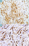
GIST. Diffuse cytoplasmatic immunopositivity for CD117 (A) and DOG1 (B) is usually observed.
Figure 5.
Figure 5.
SDH-deficient GIST. In this variant of GIST, the loss of expression of proteins forming the SDH complex (SDHB) is observed. Note the positive built-in control represented by lymphocytes and endothelial cells.
Figure 6.
Figure 6.
Desmoid fibromatosis. This tumour is composed of long fascicles of uniform, cytologically bland spindle cells, set in a collagenous background.
Figure 7.
Figure 7.
Desmoid fibromatosis. Nuclear expression of beta-catenin is a key diagnostic clue.
Figure 8.
Figure 8.
Inflammatory Myofibroblastic Tumour may rarely occur in gastrointestinal tract. In this case neoplastic cells involve the muscolaris mucosae of the small bowel.
Figure 9.
Figure 9.
Inflammatory Myofibroblastic Tumour. This tumour is composed of spindle cells, set in a fibrous stroma associated with a prominent inflammatory infiltrate, rich in plasma cells, lymphocytes and eosinophils.
Figure 10.
Figure 10.
Inflammatory Myofibroblastic Tumour. Neoplastic cells show variable positivity for smooth muscle actin (A) and in half of cases expression of ALK (B).
Figure 11.
Figure 11.
Solitary fibrous tumour. This neoplasm shows spindle cells organised in a “patternless” pattern, set in a collagenous background, and showing thin-walled, haemangiopericytoma--like vessels.
Figure 12.
Figure 12.
Solitary fibrous tumour. Expression of STAT6 represents the most specific and sensitive diagnostic marker.
Figure 13.
Figure 13.
Inflammatory fibroid polyp. The neoplasm is composed of a proliferation of bland, spindle cells, associated with an eosinophilic-rich inflammatory infiltrate.
Figure 14.
Figure 14.
Plexiform fibromyxoma. Proliferation of bland, spindle-shaped cells, set in a fibromyxoid stroma that shows a network of thin-walled capillary-size blood vessels.
Figure 15.
Figure 15.
Lipoma. The tumour is composed of mature fat tissue without atypia and variation in size of adipocytic cells. Fibrous septa and histiocytes may be sometime observed.
Figure 16.
Figure 16.
Well Differentiated Liposarcoma. The tumour is composed of atypical lipomatous proliferation with focal nuclear atypia intersected by thick fibrous septa with hyperchromatic atypical stromal spindle cells.
Figure 17.
Figure 17.
Well Differentiated Liposarcoma. Numerous lipoblasts are rarely observed in WDLPS sometimes they can be completely absent.
Figure 18.
Figure 18.
Well Differentiated Liposarcoma. Strong MDM2 nuclear positivity is consistently observed in neoplastic cells including lipoblasts.
Figure 19.
Figure 19.
Dedifferentiated liposarcoma. The dedifferentiated component is composed of spindle and pleomorphic cells set in a fibromyxoid stroma.
Figure 20.
Figure 20.
Leiomyoma. The neoplasm is composed of non-atypical, eosinophilic, spindle cells, with blunt ended nuclei.
Figure 21.
Figure 21.
Leiomyoma. Strong and diffuse immunopositivity for desmin is usually observed.
Figure 22.
Figure 22.
Leiomyoma. KIT immunostaining highlights the hyperplasia of the interstitial cells of Cajal.
Figure 23.
Figure 23.
Leiomyosarcoma. The tumour is composed of long fascicles of eosinophilic, spindle cells with blunt ended nuclei.
Figure 24.
Figure 24.
Mucosal hamartoma. The morphological feature is represented by a proliferation of bland spindle cells set in the lamina propria, featuring wavy nuclei.
Figure 25.
Figure 25.
Mucosal hamartoma. S100 immunopositivity is consistently observed.
Figure 26.
Figure 26.
Schwannoma. The tumour is composed of spindle cells organized in short fascicles surrounded by a lymphoid cuff that represents an important diagnostic clue.
Figure 27.
Figure 27.
Schwannoma. Thick-walled blood vessels are only occasionally observed in GI-tract schwannomas.
Figure 28.
Figure 28.
Schwannoma. S100 is diffusely and strongly positive.
Figura 29.
Figura 29.
Glomus tumour. This neoplasm is composed of epithelioid cells harbouring round nuclei and showing sharply delineated cytoplasm, arranged around blood vessels.
Figure 30.
Figure 30.
M-GNET. Malignant gastrointestinal neuroectodermal tumour is composed of a combination of neoplastic cells featuring eosinophilic and clear cytoplasm.
Figure 31.
Figure 31.
M-GNET. Multinucleated giant cells are relatively often seen.
Figure 32.
Figure 32.
M-GNET. Diffuse S100 immunopositivity is seen.
Figure 33.
Figure 33.
Synovial sarcoma. A primary gastric monophasic synovial sarcoma is seen.
Figure 34.
Figure 34.
Synovial sarcoma, monophasic. This highly cellular neoplasm is composed of monomorphic, atypical spindle cells.
Figure 35.
Figure 35.
Synovial sarcoma, biphasic. Epithelial component exhibits a glandular configuration.
Figure 36.
Figure 36.
Synovial sarcoma. Pancytokeratins expression in monophasyc SS (A); biphasyc SS (B). Nuclear TLE1 expression is consistently observed but tends to exhibit low specificity (C). Currently the best marker is represented by the expression of SS18-SSX fusion-specific antibody (D).

References

    1. Sbaraglia M, Dei Tos AP. The pathology of soft tissue sarcomas. Radiol Med 2019;124:266-281. https://doi.org/10.1007/s11547-018-0882-7 10.1007/s11547-018-0882-7 - DOI - PubMed
    1. Sbaraglia M, Bellan E, Dei Tos AP. The 2020 WHO Classification of Soft Tissue Tumours: news and perspectives. Pathologica 2021;113:70-84. https://doi.org/10.32074/1591-951X-213. 10.32074/1591-951X-213 Epub 2020 Nov 3. PMID: 33179614 - DOI - PMC - PubMed
    1. WHO Classification of Tumours Editorial Board. Digestive system tumours. Lyon (France): International Agency for Research on Cancer; 2019
    1. Rossi S, Gasparotto D, Toffolatti L, et al. . Molecular and clinicopathologic characterization of gastrointestinal stromal tumors (GISTs) of small size. Am J Surg Pathol 2010;34:1480-1491. https://doi.org/10.1097/PAS.0b013e3181ef7431 10.1097/PAS.0b013e3181ef7431 - DOI - PubMed
    1. Miettinen M, El-Rifai W, Sobin LHL, et al. . Evaluation of malignancy and prognosis of gastrointestinal stromal tumors: a review. Hum Pathol 2002;33:478-483. https://doi.org/10.1053/hupa.2002.124123 10.1053/hupa.2002.124123 - DOI - PubMed

MeSH terms