. 2021 Mar;17(1):200286.
doi: 10.1183/20734735.0286-2020.

Acute pulmonary function decline and radiographic abnormalities: chronic cause?

Affiliations

Acute pulmonary function decline and radiographic abnormalities: chronic cause?

Kathleen M Capaccione et al. Breathe (Sheff). 2021 Mar.

Abstract

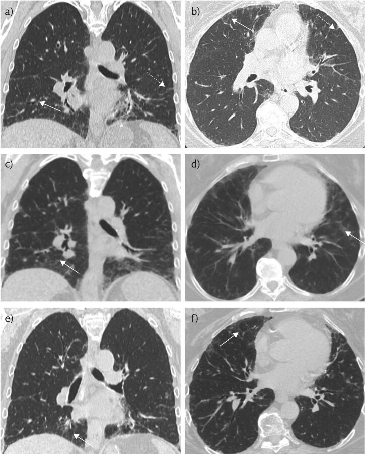
Nitrofurantoin is a cause of drug-induced pneumonitis and can result in clinically significant respiratory symptoms manifesting as interstitial lung disease on chest CT, even if the patient has been taking the drug chronically without side-effects https://bit.ly/3v2m29h.


Conflict of interest statement

Conflict of interest: K.M. Capaccione has nothing to disclose. Conflict of interest: C.V. Tran has nothing to disclose. Conflict of interest: J.S. Leb has nothing to disclose. Conflict of interest: M.M. Salvatore reports grants and other from Genentech (Speaker and Advisory Board), grants and other from Boehringer Ingelheim (Speaker and Advisory Board), outside the submitted work. Conflict of interest: B. D'souza has nothing to disclose.

Figures

Figure 1
Figure 1
a, b) Non-contrast chest CT 8 months prior to presentation, when the patient was mildly symptomatic, in the coronal and axial planes, respectively. These show mild basilar-predominant peripheral reticulations (dashed white arrows) and minimal GGOs (solid white arrows). c, d) Representative coronal and axial images during acute exacerbation, the solid white arrows indicate GGOs. e, f) Representative coronal and axial images after discontinuation of nitrofurantoin and initiation of steroid therapy, with near-complete resolution of GGOs and peripheral reticulations.
Figure 2
Figure 2
Representative a) coronal and b) axial images from a non-contrast chest CT of the abdomen and pelvis of this 64-year-old female obtained to evaluate for renal calculus. An obstructive calculus is seen in the distal right ureter (solid white arrow) with proximal hydroureteronephrosis (dotted white arrow).

References

    1. Cooper JA, Jr, White DA, Matthay RA. Drug-induced pulmonary disease. Part 2: Noncytotoxic drugs. Am Rev Respir Dis 1986; 133: 488–505. doi:10.1164/arrd.1986.133.3.488 - DOI - PubMed
    1. Rossi SE, Erasmus JJ, McAdams HP, et al. . Pulmonary drug toxicity: radiologic and pathologic manifestations. RadioGraphics 2000; 20: 1245–1259. doi:10.1148/radiographics.20.5.g00se081245 - DOI - PubMed
    1. Padley SP, Adler B, Hansell DM, et al. . High-resolution computed tomography of drug-induced lung disease. Clin Radiol 1992; 46: 232–236. doi:10.1016/S0009-9260(05)80161-8 - DOI - PubMed
    1. Goemaere NN, Grijm K, van Hal PT, et al. . Nitrofurantoin-induced pulmonary fibrosis: a case report. J Med Case Rep 2008; 2: 169. doi:10.1186/1752-1947-2-169 - DOI - PMC - PubMed
    1. Chin AJ, Rashid S, Gharibeh TR, et al. . Interstitial lung disease secondary to long-term nitrofurantoin use. Am J Case Rep 2020; 21: e920386. - PMC - PubMed

LinkOut - more resources