Review
. 2021 Jul 6:9:695900.
doi: 10.3389/fcell.2021.695900. eCollection 2021.

Mesenchymal Stem Cells in Treatment of Spinal Cord Injury and Amyotrophic Lateral Sclerosis

Affiliations
Review

Mesenchymal Stem Cells in Treatment of Spinal Cord Injury and Amyotrophic Lateral Sclerosis

Eva Sykova et al. Front Cell Dev Biol. .

Erratum in

Abstract

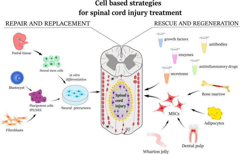
Preclinical and clinical studies with various stem cells, their secretomes, and extracellular vesicles (EVs) indicate their use as a promising strategy for the treatment of various diseases and tissue defects, including neurodegenerative diseases such as spinal cord injury (SCI) and amyotrophic lateral sclerosis (ALS). Autologous and allogenic mesenchymal stem cells (MSCs) are so far the best candidates for use in regenerative medicine. Here we review the effects of the implantation of MSCs (progenitors of mesodermal origin) in animal models of SCI and ALS and in clinical studies. MSCs possess multilineage differentiation potential and are easily expandable in vitro. These cells, obtained from bone marrow (BM), adipose tissue, Wharton jelly, or even other tissues, have immunomodulatory and paracrine potential, releasing a number of cytokines and factors which inhibit the proliferation of T cells, B cells, and natural killer cells and modify dendritic cell activity. They are hypoimmunogenic, migrate toward lesion sites, induce better regeneration, preserve perineuronal nets, and stimulate neural plasticity. There is a wide use of MSC systemic application or MSCs seeded on scaffolds and tissue bridges made from various synthetic and natural biomaterials, including human decellularized extracellular matrix (ECM) or nanofibers. The positive effects of MSC implantation have been recorded in animals with SCI lesions and ALS. Moreover, promising effects of autologous as well as allogenic MSCs for the treatment of SCI and ALS were demonstrated in recent clinical studies.

Keywords: amyotrophic lateral sclerosis; biomaterials; cell therapy; conditioned medium; exosomes; mesenchymal stem cells; neurodegenerative diseases; spinal cord injury.


Conflict of interest statement

The authors declare that the research was conducted in the absence of any commercial or financial relationships that could be construed as a potential conflict of interest.

Figures

FIGURE 1
FIGURE 1
Schematic illustration describing the strategies for the treatment of spinal cord injury. Repair and replacement of damaged tissue by neural precursors derived either from fetal neural tissue or from induced pluripotent stem cells (iPS/hES). The mesenchymal stem cells derived from bone marrow, adipose tissue, dental pulp, or umbilical cord Wharton’s jelly have a rescue-and-regeneration effect mediated by paracrine action through releasing secretomes, growth factors, anti-inflammatory molecules as well as enzymes and antibodies.
FIGURE 2
FIGURE 2
Bone marrow mesenchymal stem cells (BMSCs) labeled with iron-oxide nanoparticles implanted into rat with acute balloon-induced spinal cord compression lesion. (A,B) Longitudinal MRI images of spinal cord lesion. (A) At 5 weeks after compression the lesion was detected as a hyperintensive area with a weak hypointense signal. (B) Entire lesion populated by intravenously injected magnetically labeled BMSCs at 4 weeks after implantation is visible as a dark hypointensive area. (C) Prussian blue staining for iron of a spinal cord lesion in control animal. (D) Prussian blue staining for iron of a spinal cord lesion at 4 weeks after labeled BMSCs implantation. Note the smaller lesion size in the animal with implanted BMSC. (E) Prussian blue staining in detail shows a staining for hemoglobin. (F) The lesion is populated with Prussian blue-positive cells. Modified from Jendelová et al. (2004).
FIGURE 3
FIGURE 3
Hematoxylin–eosin staining of a rat spinal cord with acute balloon-induced spinal cord compression lesion. Spinal cord injury (SCI) only: longitudinal section of the spinal cord with a cavity in the control animal at 6 months after SCI. SCI + hydrogel + mesenchymal stem cells (MSCs): spinal cord at 6 months after a lesion with MSC-seeded HPMA-arginine–glycine–aspartic acid hydrogel implanted at 5 weeks after SCI. Motor test and sensory test: A comparison of motor test (Basso, Beattie, and Bresnahan score) and sensory test (plantar test) in 1–25 weeks after SCI in rats with SCI only, SCI and hydrogel implantation, and SCI with implantation of hydrogel seeded with bone marrow mesenchymal stem cells. Modified from Hejcl et al. (2010).
FIGURE 4
FIGURE 4
(A–C) SEM micrographs of (A) extracellular matrix (ECM) hydrogel, (B) hyaluronic acid (HA) hydrogel modified with arginine–glycine–aspartic acid (RGD), and (C) highly superporous SIKVAV-modified superporous poly (2-hydroxyethyl methacrylate) hydrogel scaffolds with oriented pores. (D–I) Representative images of the longitudinal sections of the spinal cord lesion after hydrogel injection or implantation into the hemisection cavity. (D,E) Immunofluorescence staining for neurofilaments (NF-160, green) and (E) cell nuclei (DAPI, blue) at 2 weeks after the injection of ECM hydrogel derived from porcine spinal cord. (F,G) Immunofluorescence staining for neurofilaments (NF-160, green), (G) astrocytes (GFAP, red), and cell nuclei (DAPI, blue) at 8 weeks after HA–RGD hydrogel implantation; the square in panel (F) is shown under the higher magnification inset in panel (G). (H,I) Immunofluorescence staining for (H) blood vessels (RECA) and (I) neurofilaments (NF-160, green) at 2 months after the implantation of SIKVAV-modified superporous poly (2-hydroxyethyl methacrylate) hydrogel with parallel-oriented pores. Scale bar: (D,F,H) 500 μmm, (E,G) 50 μmm, and (I) 100 μmm. Modified from (A) Koci et al. (2017), (D,E) Tukmachev et al. (2016), (B,F,G) Zaviskova et al. (2018), and (C,H,I) Kubinova et al. (2015).
FIGURE 5
FIGURE 5
Clinical analysis of 12 amyotrophic lateral sclerosis patients with fast decline of functional rating scale (ALSFRS) scores 6 months before and 12 months after autologous bone marrow mesenchymal stem cell (BMSC) application. The upper panel shows the time courses of ALSFRS scores in individual patients; patient no. 1 is shown in the inset. The lower panel shows the regression analysis of ALSFRS scores before and after BMSC application. The solid line with β = –0.80 is the predicted time course without BMSC treatment. Modified from Sykova et al. (2017).

References

    1. Ahmadian Kia N., Bahrami A. R., Ebrahimi M., Matin M. M., Neshati Z., Almohaddesin M. R., et al. (2011). Comparative analysis of chemokine receptor’s expression in mesenchymal stem cells derived from human bone marrow and adipose tissue. J. Mol. Neurosci. 44 178–185. 10.1007/s12031-010-9446-6 - DOI - PubMed
    1. Ahuja C. S., Wilson J. R., Nori S., Kotter M. R. N., Druschel C., Curt A., et al. (2017). Traumatic spinal cord injury. Nat. Rev. Dis. Primers. 3:17018. - PubMed
    1. Amemori T., Ruzicka J., Romanyuk N., Jhanwar-Uniyal M., Sykova E., Jendelova P. (2015). Comparison of intraspinal and intrathecal implantation of induced pluripotent stem cell-derived neural precursors for the treatment of spinal cord injury in rats. Stem Cell. Res. Ther. 6:257. - PMC - PubMed
    1. Angeli C. A., Boakye M., Morton R. A., Vogt J., Benton K., Chen Y., et al. (2018). Recovery of over-ground walking after chronic motor complete spinal cord injury. N. Engl. J. Med. 379 1244–1250. 10.1056/nejmoa1803588 - DOI - PubMed
    1. Asadi-Golshan R., Razban V., Mirzaei E., Rahmanian A., Khajeh S., Mostafavi-Pour Z., et al. (2018). Sensory and motor behavior evidences supporting the usefulness of conditioned medium from dental pulp-derived stem cells in spinal cord injury in rats. Asian Spine J. 12 785–793. 10.31616/asj.2018.12.5.785 - DOI - PMC - PubMed