

Behavioral and Neural Signatures of Visual Imagery Vividness Extremes: Aphantasia versus Hyperphantasia
- PMID: 34296179
- PMCID: PMC8186241
- DOI: 10.1093/texcom/tgab035
Behavioral and Neural Signatures of Visual Imagery Vividness Extremes: Aphantasia versus Hyperphantasia
Abstract
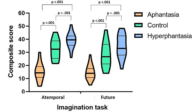
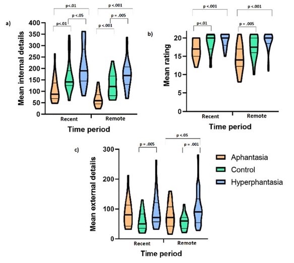
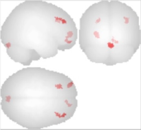
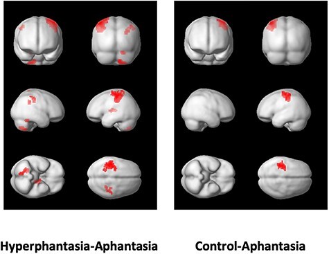
Although Galton recognized in the 1880s that some individuals lack visual imagery, this phenomenon was mostly neglected over the following century. We recently coined the terms "aphantasia" and "hyperphantasia" to describe visual imagery vividness extremes, unlocking a sustained surge of public interest. Aphantasia is associated with subjective impairment of face recognition and autobiographical memory. Here we report the first systematic, wide-ranging neuropsychological and brain imaging study of people with aphantasia (n = 24), hyperphantasia (n = 25), and midrange imagery vividness (n = 20). Despite equivalent performance on standard memory tests, marked group differences were measured in autobiographical memory and imagination, participants with hyperphantasia outperforming controls who outperformed participants with aphantasia. Face recognition difficulties and autistic spectrum traits were reported more commonly in aphantasia. The Revised NEO Personality Inventory highlighted reduced extraversion in the aphantasia group and increased openness in the hyperphantasia group. Resting state fMRI revealed stronger connectivity between prefrontal cortices and the visual network among hyperphantasic than aphantasic participants. In an active fMRI paradigm, there was greater anterior parietal activation among hyperphantasic and control than aphantasic participants when comparing visualization of famous faces and places with perception. These behavioral and neural signatures of visual imagery vividness extremes validate and illuminate this significant but neglected dimension of individual difference.
Keywords: aphantasia; autobiographical; hyperphantasia; imagery; neuroimaging.
© The Author(s) 2021. Published by Oxford University Press.
Figures
References
-
- Andersson JLR, Jenkinson M, Smith S. 2007a. Non-linear Optimisation. FMRIB technical report TR07JA1. www.fmrib.ox.ac.uk/analysis/techrep.
-
- Andersson JLR, Jenkinson M, Smith S. 2007b. Non-linear Registration, AKA Spatial Normalisation. FMRIB technical report TR07JA2. www.fmrib.ox.ac.uk/analysis/techrep.
-
- Ashburner J. 2007. A fast diffeomorphic image registration algorithm. NeuroImage. 38(1):95–113. - PubMed
LinkOut - more resources
Full Text Sources
Other Literature Sources