. 2022 Jan 5;24(1):52-63.
doi: 10.1093/neuonc/noab177.

Selective blood-brain barrier permeabilization of brain metastases by a type 1 receptor-selective tumor necrosis factor mutein

Affiliations

Selective blood-brain barrier permeabilization of brain metastases by a type 1 receptor-selective tumor necrosis factor mutein

Mario F Munoz Pinto et al. Neuro Oncol. .

Abstract

Background: Metastasis to the brain is a major challenge with poor prognosis. The blood-brain barrier (BBB) is a significant impediment to effective treatment, being intact during the early stages of tumor development and heterogeneously permeable at later stages. Intravenous injection of tumor necrosis factor (TNF) selectively induces BBB permeabilization at sites of brain micrometastasis, in a TNF type 1 receptor (TNFR1)-dependent manner. Here, to enable clinical translation, we have developed a TNFR1-selective agonist variant of human TNF that induces BBB permeabilization, while minimizing potential toxicity.

Methods: A library of human TNF muteins (mutTNF) was generated and assessed for binding specificity to mouse and human TNFR1/2, endothelial permeabilizing activity in vitro, potential immunogenicity, and circulatory half-life. The permeabilizing ability of the most promising variant was assessed in vivo in a model of brain metastasis.

Results: The primary mutTNF variant showed similar affinity for human TNFR1 than wild-type human TNF, similar affinity for mouse TNFR1 as wild-type mouse TNF, undetectable binding to human/mouse TNFR2, low potential immunogenicity, and permeabilization of an endothelial monolayer. Circulatory half-life was similar to mouse/human TNF and BBB permeabilization was induced selectively at sites of micrometastases in vivo, with a time window of ≥24 hours and enabling delivery of agents within a therapeutically relevant range (0.5-150 kDa), including the clinically approved therapy, trastuzumab.

Conclusions: We have developed a clinically translatable mutTNF that selectively opens the BBB at micrometastatic sites, while leaving the rest of the cerebrovasculature intact. This approach will open a window for brain metastasis treatment that currently does not exist.

Keywords: blood-brain barrier; brain metastasis; mutein; permeabilization; tumor necrosis factor.


Figures

Fig. 1
Fig. 1
Biological activity of mutTNF G4 in vitro. (A) Single-cycle kinetic sensorgram data (red lines) and fitted curves (black lines) for the binding of huTNF and mutTNF G4 to huTNFR1, huTNFR2, mTNFR1, and mTNFR2. (B) Assessment of alterations in permeability as reflected by changes in permeability coefficients (Pe) of a hCMEC/D3 cell monolayer treated with 0.1-10 ng/mL of huTNF (WT) or mutTNF (G4) for 24 hours, VEGF (V) as positive control, or untreated cells (U) as negative control. (C) Plasma concentrations (ng/mL) for mutTNF G4 at different time points after intravenous administration of 1 µg. Statistical analysis: all values are expressed as mean ± SD. 1-way ANOVA with Sidak post hoc test vs untreated group (***P < .005) or huTNF group (##P < .01). Abbreviations: mutTNF, TNF muteins; VEGF, vascular endothelial growth factor.
Fig. 2
Fig. 2
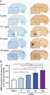
Histological assessment of mutTNF-induced BBB breakdown by HRP histochemistry. (A) Hanker-Yates histology for HRP (44 kDa) detection (brown) revealed areas of BBB breakdown at metastatic sites, as confirmed by cresyl violet histology on adjacent sections. (B) Graph showing dose-response analysis of metastasis-specific BBB breakdown frequency 2 hours after different doses of mutTNF (n = 3/group, except 50 µg/kg and saline where n = 4). Scale bars = 200 µm (enlarged inner square) and 1 mm. Statistical analysis: all values are expressed as mean ± SD. 1-way ANOVA with post hoc Tukey test (*P < .05, ***P < .005). Abbreviations: BBB, blood-brain barrier; HRP, horseradish peroxidase; mutTNF, TNF muteins.
Fig. 3
Fig. 3
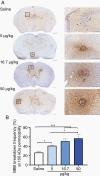
Histological assessment of mutTNF-induced BBB breakdown by IgG immunostaining. (A) IgG (150 kDa) immunostaining (brown) revealed areas of BBB breakdown at metastatic sites; sections counterstained with cresyl violet. (B) Graph showing dose-response analysis of metastasis-specific BBB breakdown frequency 2 hours after different doses of mutTNF (n = 3/group). Scale bars = 200 µm (enlarged inner square) and 1 mm. Statistical analysis: all values are expressed as mean ± SD. 1-way ANOVA with post hoc Tukey test (*P < .05, ***P < .005). Abbreviations: BBB, blood-brain barrier; mutTNF, TNF muteins.
Fig. 4
Fig. 4
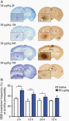
Time-course of mutTNF-induced BBB breakdown. (A, B) Hanker-Yates histology for HRP detection (brown) revealed areas of BBB breakdown at metastatic sites, as confirmed by cresyl violet histology on adjacent sections, at different time points in saline (A) and mutTNF G4 (B) treated animals. (C) Graph showing frequency of metastasis-specific BBB breakdown at different time points after systemic administration of 50 µg/kg mutTNF (n = 6/group for mutTNF-treated animals; n = 3/group for saline-treated animals). Scale bars = 200 µm (enlarged inner square) and 1 mm. Statistical analysis: all values are expressed as mean ± SD. 1-way ANOVA with Sidak post hoc test vs saline group (*P < .05, ***P < .005). Abbreviations: BBB, blood-brain barrier; HRP, horseradish peroxidase; mutTNF, TNF muteins.
Figure 5.
Figure 5.
Gd-DTPA enhancement at metastatic sites. T1-weighted images were acquired pre- (A, C) and 5-minute post-Gd-DTPA injection (B, D) 2 hours after systemic administration of 16.7 µg/kg (B) or 5 µg/kg (D) of mutTNF G4. Areas of high signal intensity (white) infer areas of BBB breakdown. The presence of metastasis within the white box in (B) was confirmed on a cresyl violet stained section (E) together with the adjacent Hanker-Yates section (F) confirming HRP extravasation (brown staining), and hence, BBB breakdown. (G) Histological confirmation of the presence of metastatic foci within the white box in (D) on a corresponding IgG-stained section; a metastasis is clearly visible at the center of the brown-stained region in the zoomed image of the boxed region in (H). Sections counterstained with cresyl violet. Scale bars = 200 µm (enlarged inner square) and 1 mm. (I) Graph showing percentage increase in signal intensity at metastatic sites (n = 6/group). Statistical analysis: all values are expressed as mean ± SD. Nonparametric Kruskal-Wallis test was performed with Dunn post hoc test vs saline group (*P < .05, **P < .01, ***P < .005). Abbreviations: BBB, blood-brain barrier; HRP, horseradish peroxidase.
Figure 6.
Figure 6.
Trastuzumab extravasation after co-administration with either mutTNF or saline. (A–H) Example images of metastatic sites classified as either positive or negative for trastuzumab, as determined by human IgG immunostaining (brown). (A–D) Examples of IgG-positive metastases in a saline-treated mouse (A, B) and a mutTNF G4-treated mouse (C, D). (E–H) Examples of IgG negative metastases in a saline-treated mouse (E, F) and a mutTNF G4-treated mouse (G, H). Sections were first assessed for trastuzumab positivity (B, D, F, H; brown staining), and subsequently, counterstained with cresyl violet to verify the presence of metastases (A, C, G, E). Scale bars = 150 µm. (I) Graph showing quantitative analysis of trastuzumab extravasation at sites of metastasis from saline control (n = 3 mice; n = 714 metastases) and mutTNF-treated (n = 3 mice; n = 978 metastases) groups; significantly greater extravasation of trastuzumab was evident in mutTNF-treated animals. Statistical analysis: all values are expressed as mean ± SD. Nonparametric Mann-Whitney U test (**P < .01). Abbreviation: mutTNF, TNF muteins.

References

    1. Eichler AF, Chung E, Kodack DP, Loeffler JS, Fukumura D, Jain RK. The biology of brain metastases-translation to new therapies. Nat Rev Clin Oncol. 2011;8(6):344–356. - PMC - PubMed
    1. Hersh DS, Wadajkar AS, Roberts N, et al. . Evolving drug delivery strategies to overcome the blood brain barrier. Curr Pharm Des. 2016;22(9):1177–1193. - PMC - PubMed
    1. Côté J, Bovenzi V, Savard M, et al. . Induction of selective blood-tumor barrier permeability and macromolecular transport by a biostable kinin B1 receptor agonist in a glioma rat model. PLoS One. 2012;7(5):e37485. - PMC - PubMed
    1. Côté J, Savard M, Neugebauer W, Fortin D, Lepage M, Gobeil F. Dual kinin B1 and B2 receptor activation provides enhanced blood-brain barrier permeability and anticancer drug delivery into brain tumors. Cancer Biol Ther. 2013;14(9):806–811. - PMC - PubMed
    1. Guillaume DJ, Doolittle ND, Gahramanov S, Hedrick NA, Delashaw JB, Neuwelt EA. Intra-arterial chemotherapy with osmotic blood-brain barrier disruption for aggressive oligodendroglial tumors: results of a phase I study. Neurosurgery. 2010;66(1):48–58; discussion 58. - PMC - PubMed

Publication types