Review
. 2021 Nov;63(11):1765-1772.
doi: 10.1007/s00234-021-02766-z. Epub 2021 Jul 23.

Spontaneous intracranial hypotension: diagnostic and therapeutic workup

Affiliations
Review

Spontaneous intracranial hypotension: diagnostic and therapeutic workup

Niklas Luetzen et al. Neuroradiology. 2021 Nov.

Abstract

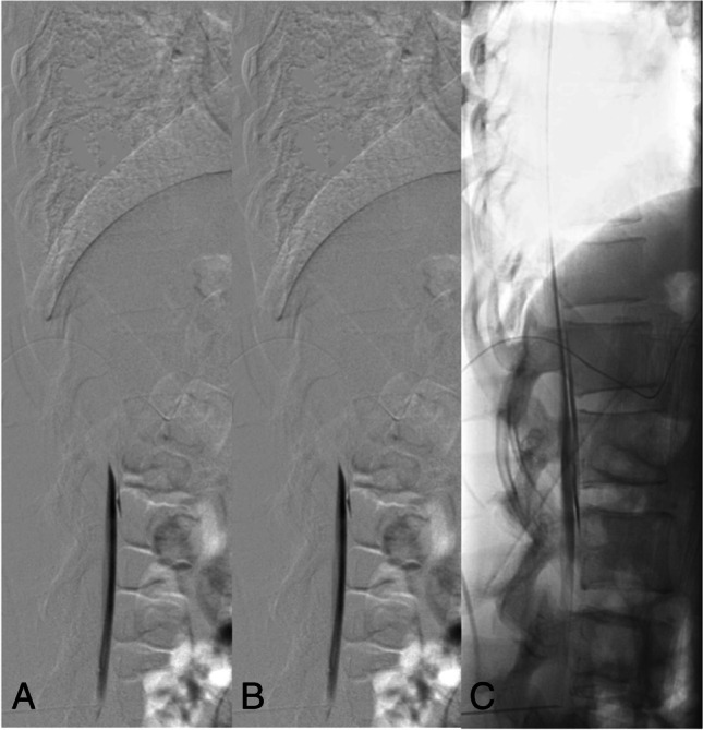
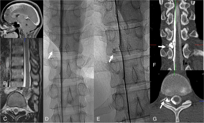
Spontaneous intracranial hypotension (SIH) is an orthostatic headache syndrome with typical MRI findings among which engorgement of the venous sinuses, pachymeningeal enhancement, and effacement of the suprasellar cistern have the highest diagnostic sensitivity. SIH is in almost all cases caused by spinal CSF leaks. Spinal MRI scans showing so-called spinal longitudinal extradural fluid (SLEC) are suggestive of ventral dural tears (type 1 leak) which are located with prone dynamic (digital subtraction) myelography. As around half of the ventral dural tears are located in the upper thoracic spine, additional prone dynamic CT myelography is often needed. Leaking nerve root sleeves typically associated with meningeal diverticulae (type 2 leaks) and CSF-venous fistulas (type 3 leaks) are proven via lateral decubitus dynamic digital subtraction or CT myelography: type 2 leaks are SLEC-positive if the tear is proximal and SLEC-negative if it is distal, and type 3 leaks are always SLEC-negative. Although 30-70% of SIH patients show marked improvement following epidural blood patches applied via various techniques definite cure mostly requires surgical closure of ventral dural tears and surgical ligations of leaking nerve root sleeves associated with meningeal diverticulae or CSF-venous fistulas. For the latter, transvenous embolization with liquid embolic agents via the azygos vein system is a novel and valuable therapeutic alternative.

Keywords: CSF-venous fistula; Lateral decubitus myelography; Spontaneous intracranial hypotension.


Conflict of interest statement

The authors declare that they have no conflict of interest.

Figures

Fig. 1
Fig. 1
Diagnostic algorithm for patients with suspected SIH
Fig. 2
Fig. 2
Type 1 leak: Dynamic digital subtraction myelogram (DSM) with the patient in prone position on a tilted table. Due to the higher specific weight compared to CSF, iodinated contrast medium collects at the ventral surface of the thecal sac. With the X-ray tube in lateral and the patient in head down position (around 15°), digital subtraction myelograms are acquired during gentle injection of highly concentrated (300 mg/ml) contrast medium. Using this technique, most of the ventral leaks in the lower thoracic and lumbar spine can be visualized. In this 30-year-old woman, dynamic DSM shows a ventral leak at the level L1/2 (A, B). Note that on the subsequent unsubtracted fluoroscopic image the exact site of the leak cannot be determined any longer (C)
Fig. 3
Fig. 3
Type 1 leak: SIH in a 36-year-old woman with engorgement of the venous sinuses, pachymeningeal enhancement (B: arrow), subdural fluid collections (B: hollow arrow), effaced suprasellar (C: 1.5 mm), and prepontine cisterns (C: 2.7 mm). The mamillopontine distance is normal (C: 7.3 mm). Spinal MRI shows spinal longitudinal extradural fluid (SLEC) (D, E: hollow arrow). Prone dynamic CT myelography shows the ventral leak due to a bony spur (H: arrow) and ventral epidural contrast flowing upwards (F, G: hollow arrow)
Fig. 4
Fig. 4
Type 2 leak: Right-sided lateral decubitus myelography shows a leaking meningeal diverticulum Th 10/11 on the right-side (A). Subsequent lateral decubitus CT myelography (B, C) shows extradural contrast (B: arrow) indicating a proximal leak
Fig. 5
Fig. 5
Type 2 leak according to Schievink or type 4 leak according to Farb, respectively: Head-positive (A), SLEC-negative MRI scans (B, C). Right-sided lateral decubitus dynamic myelography shows a slowly filling meningeal diverticulum (D, E: arrow). Right-sided lateral decubitus dynamic CT myelography shows the extrathecal contrast (F: G: arrow)
Fig. 6
Fig. 6
Type 3 leak: Right-sided lateral decubitus dynamic myelography shows a CSF-venous fistula, i.e., here, the tiny venous filling at the level Th 10/11 (A: arrow). Right-sided lateral decubitus CT myelography clearly shows a hyperdense paraspinal vein confirming a CSF-venous fistula (B: arrow). For transvenous embolization, a Scepter dual‐lumen balloon catheter (Microvention, Tustin, CA, USA) is navigated via the azygos vein into the collector of the veins surrounding the nerve root (C: The position of the nerve root is marked with an arrow). Onyx deposition (D). Post-embolization CT shows Onyx cast in the desired position (E, F)

References

    1. Headache Classification Committee of the International Headache Society (IHS) (2018) The international classification of headache disorders, 3rd edition. Cephalalgia 38:1–211 - PubMed
    1. Schievink WI, Maya MM, Moser F, Tourje J, Torbati S. Frequency of spontaneous intracranial hypotension in the emergency department. J Headache Pain. 2007;8:325–328. doi: 10.1007/s10194-007-0421-8. - DOI - PMC - PubMed
    1. Schievink WI, Meyer FB, Atkinson JLD, Mokri B. Spontaneous spinal cerebrospinal fluid leaks and intracranial hypotension. J Neurosurg. 1996;84:598–605. doi: 10.3171/jns.1996.84.4.0598. - DOI - PubMed
    1. Schievink WI, Morreale VM, Atkinson JL, Meyer FB, Piepgras DG, Ebersold MJ. Surgical treatment of spontaneous spinal cerebrospinal fluid leaks. J Neurosurg. 1998;88:243–246. doi: 10.3171/jns.1998.88.2.0243. - DOI - PubMed
    1. Schievink WI. Spontaneous spinal cerebrospinal fluid leaks and intracranial hypotension. JAMA. 2006;295:2286–2295. doi: 10.1001/jama.295.19.2286. - DOI - PubMed

MeSH terms