

A multicentre and multi-national evaluation of the accuracy of quantitative Lu-177 SPECT/CT imaging performed within the MRTDosimetry project
- PMID: 34297218
- PMCID: PMC8302709
- DOI: 10.1186/s40658-021-00397-0
A multicentre and multi-national evaluation of the accuracy of quantitative Lu-177 SPECT/CT imaging performed within the MRTDosimetry project
Abstract
Purpose: Patient-specific dosimetry is required to ensure the safety of molecular radiotherapy and to predict response. Dosimetry involves several steps, the first of which is the determination of the activity of the radiopharmaceutical taken up by an organ/lesion over time. As uncertainties propagate along each of the subsequent steps (integration of the time-activity curve, absorbed dose calculation), establishing a reliable activity quantification is essential. The MRTDosimetry project was a European initiative to bring together expertise in metrology and nuclear medicine research, with one main goal of standardizing quantitative 177Lu SPECT/CT imaging based on a calibration protocol developed and tested in a multicentre inter-comparison. This study presents the setup and results of this comparison exercise.
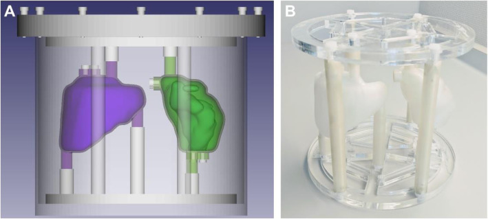
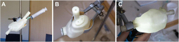
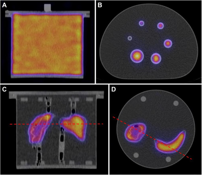
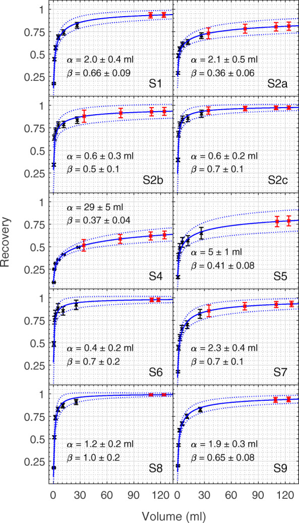
Methods: The inter-comparison included nine SPECT/CT systems. Each site performed a set of three measurements with the same setup (system, acquisition and reconstruction): (1) Determination of an image calibration for conversion from counts to activity concentration (large cylinder phantom), (2) determination of recovery coefficients for partial volume correction (IEC NEMA PET body phantom with sphere inserts), (3) validation of the established quantitative imaging setup using a 3D printed two-organ phantom (ICRP110-based kidney and spleen). In contrast to previous efforts, traceability of the activity measurement was required for each participant, and all participants were asked to calculate uncertainties for their SPECT-based activities.
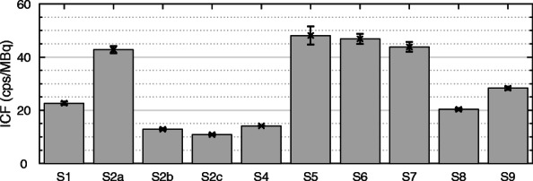
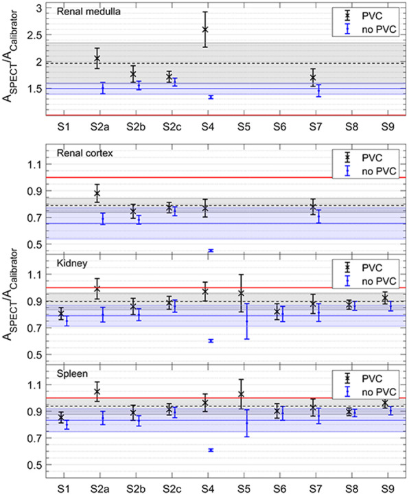
Results: Similar combinations of imaging system and reconstruction lead to similar image calibration factors. The activity ratio results of the anthropomorphic phantom validation demonstrate significant harmonization of quantitative imaging performance between the sites with all sites falling within one standard deviation of the mean values for all inserts. Activity recovery was underestimated for total kidney, spleen, and kidney cortex, while it was overestimated for the medulla.
Conclusion: This international comparison exercise demonstrates that harmonization of quantitative SPECT/CT is feasible when following very specific instructions of a dedicated calibration protocol, as developed within the MRTDosimetry project. While quantitative imaging performance demonstrates significant harmonization, an over- and underestimation of the activity recovery highlights the limitations of any partial volume correction in the presence of spill-in and spill-out between two adjacent volumes of interests.
Keywords: 177Lu SPECT/CT imaging; 3D printing; Harmonization of SPECT/CT imaging; International multicenter comparison exercise; Molecular radiotherapy (MRT); Phantom; Quantitative SPECT/CT; Standardization of SPECT/CT imaging; Traceability of SPECT/CT imaging.
© 2021. The Author(s).
Conflict of interest statement
M. Lassmann has received research grants by IPSEN Pharma and Nordic Nanovector. M. Ljungberg and K. Sjögreen-Gleisner have been acting as research consultants for Fusion Pharmaceuticals Inc, Canada.
Figures
References
-
- Dewaraja YK, Frey EC, Sgouros G, Brill AB, Roberson P, Zanzonico PB, Ljungberg M. MIRD pamphlet No. 23: quantitative SPECT for patient-specific 3-dimensional dosimetry in internal radionuclide therapy. Journal of nuclear medicine : official publication. Soc Nuclear Med. 2012;53(8):1310–1325. doi: 10.2967/jnumed.111.100123. - DOI - PMC - PubMed
Grants and funding
LinkOut - more resources
Full Text Sources
Medical
Research Materials
Miscellaneous
