

doi: 10.1111/bpa.13008.
Epub 2021 Jul 23.
Myxoid glioneuronal tumor, PDGFRA p.K385L-mutant, arising in midbrain tectum with multifocal CSF dissemination
Affiliations
- PMID: 34297434
- PMCID: PMC8713525
- DOI: 10.1111/bpa.13008
Item in Clipboard
Myxoid glioneuronal tumor, PDGFRA p.K385L-mutant, arising in midbrain tectum with multifocal CSF dissemination
Brain Pathol.
2022 Jan.
Abstract
This myxoid glioneuronal tumor, PDGFRA p.K385L-mutant, arose in the midbrain tectum rather than in the septum pellucidum, as in the previously-reported cases.
Keywords: DNA methylation; brainstem; glioneuronal; next generation sequencing; tumors.
© 2021 The Authors. Brain Pathology published by John Wiley & Sons Ltd on behalf of International Society of Neuropathology.
Conflict of interest statement
The author(s) declared no potential conflicts of interest with respect to the research, authorship, and/or publication of this article.
Figures
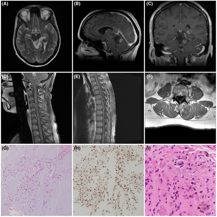
(A) Axial non‐contrast‐enhanced T2‐weighted MRI demonstrates the tectal plate origin (arrowhead) of the tumor. (B) Sagittal contrast‐enhanced T1‐weighted MRI proves the absence of corpus callosal or lateral ventricular involvement but highlights the enhancing component within the tectal tumor (arrowhead). (C) Coronal contrast‐enhanced T1‐weighted MRI shows the nodular posterior fossa spread (black arrowhead) as well as leptomeningeal enhancement within cerebellar folia (white arrowheads). (D) Sagittal contrast‐enhanced T1‐weighted MRI of cervical spine shows nodular enhancing masses over the dorsum of the cord (arrowheads). (E). Sagittal contrast‐enhanced T1‐weighted MRI of thoracic spine shows nodular enhancing masses over the dorsum of cord (arrowheads). (F) Axial post contrast T1‐weighted MRI shows nodular enhancement (arrowhead) involving the left L5 nerve roots. (G) Low power view of the small oligodendroglial‐like cells embedded in copious mucin (hematoxylin & eosin). (H) Tumor nuclei were diffusely immunoreactive for OLIG2 (IHC for OLIG2 with light hematoxylin counterstain). (I) Hemosiderin (arrowheads) and eosinophilic granular bodies (arrows) were focally identified (H&E)
References
-
- Kurtkaya‐Yapicier O, Elmaci I, Boran B, Kiliç T, Sav A, Pamir MN. Dysembryoplastic neuroepithelial tumor of the midbrain tectum: a case report. Brain Tumor Pathol. 2002;19(2):97–100. - PubMed
-
- Whittle IR, Dow GR, Lammie GA, Wardlaw J. Dsyembryoplastic neuroepithelial tumour with discrete bilateral multifocality: further evidence for a germinal origin. Br J Neurosurg. 1999;13(5):508–11. - PubMed
-
- Leung SY, Gwi E, Ng HK, Fung CF, Yam KY. Dysembryoplastic neuroepithelial tumor. A tumor with small neuronal cells resembling oligodendroglioma. Am J Surg Pathol. 1994;18(6):604–14. - PubMed
Publication types
MeSH terms
LinkOut - more resources
Full Text Sources
Medical
Miscellaneous
