Review
. 2021 Jul 22;184(15):3852-3872.
doi: 10.1016/j.cell.2021.06.024.

Fibroblasts: Origins, definitions, and functions in health and disease

Affiliations
Review

Fibroblasts: Origins, definitions, and functions in health and disease

Maksim V Plikus et al. Cell. .

Abstract

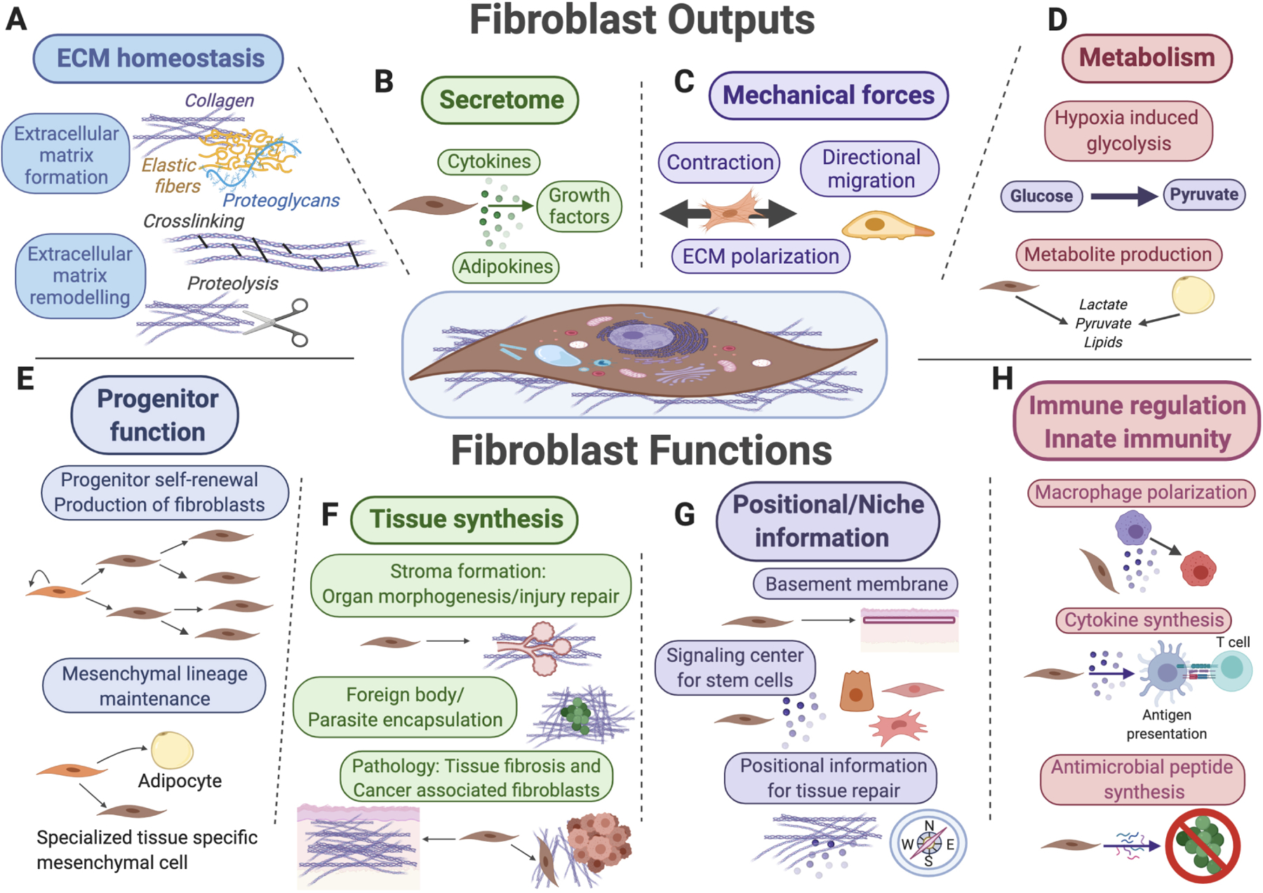
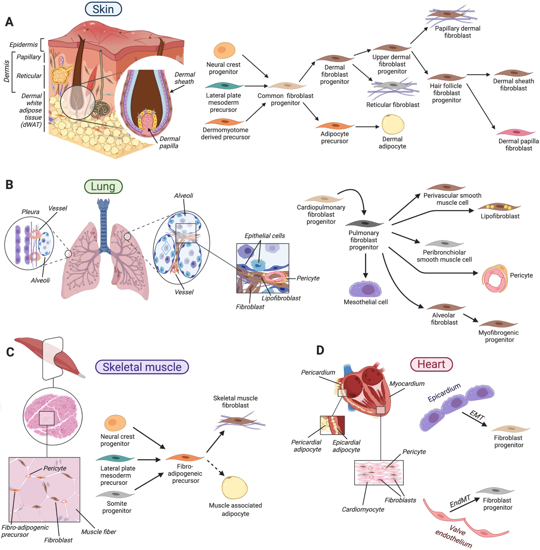
Fibroblasts are diverse mesenchymal cells that participate in tissue homeostasis and disease by producing complex extracellular matrix and creating signaling niches through biophysical and biochemical cues. Transcriptionally and functionally heterogeneous across and within organs, fibroblasts encode regional positional information and maintain distinct cellular progeny. We summarize their development, lineages, functions, and contributions to fibrosis in four fibroblast-rich organs: skin, lung, skeletal muscle, and heart. We propose that fibroblasts are uniquely poised for tissue repair by easily reentering the cell cycle and exhibiting a reversible plasticity in phenotype and cell fate. These properties, when activated aberrantly, drive fibrotic disorders in humans.


Conflict of interest statement

Declaration of interests The authors declare no competing interests.

Figures

Figure 1.
Figure 1.. Summary of fibroblast outputs and functions.
Key functions for fibroblasts (shown in the center) and their mesenchymal lineages include extracellular matrix (ECM) secretion and remodeling (A), secretion of signaling factors for surrounding cells (B), mechanical force generation (C), and regulation of tissue metabolism and metabolite secretion (D). Fibroblasts also function as progenitor cells for mesenchymal lineages (E), as “makers” of new tissue during organ morphogenesis, tissue repair and upon various pathological conditions (F), as sources of positional information across distinct anatomical regions of the same organ and as key signal contributors toward stem cell niches (G), as well as target cells and reciprocal modulators of diverse innate and adaptive immune functions (H).
Figure 2.
Figure 2.. History of fibroblast discovery.
A. First drawing of fibroblasts by Rudolf Virchow as “spindle-shaped” cells embedded within the connective tissue of pig embryo. Modified from Virchow (1858). B. Drawing by Ernst Ziegler, who first proposed the term “fibroblast” to describe cells that produce new connective tissue upon healing. Various forms of cells in the new granulation tissue are shown. Mononuclear fibroblast-shaped cells are in the bottom left corner. Modified from Ziegler (1895). C. Drawing of fibroblasts as fusiform cells within newly formed connective tissue of a “painful” keloid. Modified from Cajal (1896). D. Microphotograph of fibroblasts established from embryonic chick heart explant. Cells after 75 passages are shown. Modified from Ebeling (1913).
Figure 3.
Figure 3.. Key roles of PDGF, TGFβ and WNT signaling pathways in regulating fibroblast functions.
Platelet derived growth factor (PDGF) signaling (blue) regulates diverse aspects of fibroblast development and homeostasis, including epithelial-to-mesenchymal transition (EMT) in the embryonic heart, long-term self-renewal, and proliferation in adult tissues. Signaling via the transforming growth factor β (TGFβ) superfamily of ligands promotes myofibroblast state activation, including contractile protein and extracellular matrix (ECM) gene expression. Among other effects, TGFβ superfamily signaling can induce fibroblast proliferation and lineage transitions by other cells toward a fibroblast state, including via EMT in the lung. WNT signaling regulates fibroblast proliferation, migration, myofibroblast state activation, and ECM deposition. All three pathways can activate transcription of genes to control fibroblast biology and TGFβ activates Akt and RhoA to induce cellular contraction.
Figure 4.
Figure 4.. Diverse cellular sources of myofibroblasts.
Diverse tissue resident mesenchymal cells, including specialized fibroblast progenitors, pericytes and adipocytes can become activated and undergo reversible conversion toward a myofibroblast state. Examples of lung, skeletal muscle and skin are provided.
Figure 5.
Figure 5.. Organ-specific fibroblast organization and lineage relationships.
A. In the skin, diverse fibroblast types reside within its dermal layers, in dermal white adipose tissue and in association with hair follicles. During skin development, a common mesenchymal progenitor gives rise to dermal fibroblast progenitors that further specify toward fibroblasts of papillary and reticular dermis and hair follicle-associated fibroblasts, and to adipocyte precursors of dermal adipose tissue. B. In the lung, diverse fibroblasts associate with alveoli at the end of the branched epithelium, bronchioles and vasculature. During lung development, common cardiopulmonary progenitors generate a diverse array of fibroblasts, including mesothelial cells of the pleura, alveolar fibroblasts that support gas-exchanging epithelium, lipid droplet-containing lipofibroblasts, peribronchiolar and perivascular smooth muscle cells. C. Skeletal muscle fibroblasts, fibro-adipogenic precursors, and pericytes lie in the space between muscle fibers. Several developmentally distinct embryonic mesenchymal progenitors give rise to fibro-adipogenic precursors that, in turn, serve as long-lasting sources for muscle-associated fibroblasts as well as adipocytes upon aging and diseased states. D. Cardiac fibroblasts and pericytes reside between cardiomyocytes. During development, cardiac fibroblasts form from epicardial and endocardial epithelial cells via epithelial-to-mesenchymal transition (EMT) and endothelial-to-mesenchymal transition (EndMT), respectively. In A-D arrows indicate lineage relationships.

References

    1. Abbasi S, Sinha S, Labit E, Rosin NL, Yoon G, Rahmani W, Jaffer A, Sharma N, Hagner A, Shah P. et al. (2020). Distinct Regulatory Programs Control the Latent Regenerative Potential of Dermal Fibroblasts during Wound Healing. Cell Stem Cell 27, 396–412 e396. - PubMed
    1. Al Alam D, El Agha E, Sakurai R, Kheirollahi V, Moiseenko A, Danopoulos S, Shrestha A, Schmoldt C, Quantius J, Herold S. et al. (2015). Evidence for the involvement of fibroblast growth factor 10 in lipofibroblast formation during embryonic lung development. Development 142, 4139–4150. - PMC - PubMed
    1. Armulik A, Genove G, and Betsholtz C (2011). Pericytes: developmental, physiological, and pathological perspectives, problems, and promises. Dev Cell 21, 193–215. - PubMed
    1. Arora R, Metzger RJ, and Papaioannou VE (2012). Multiple roles and interactions of Tbx4 and Tbx5 in development of the respiratory system. PLoS Genet 8, e1002866. - PMC - PubMed
    1. Ascension AM, Fuertes-Alvarez S, Ibanez-Sole O, Izeta A, and Arauzo-Bravo MJ (2020). Human dermal fibroblast subpopulations are conserved across single-cell RNA sequencing studies. J Invest Dermatol. - PubMed

Publication types