. 2021 Jul 9;13(14):3441.
doi: 10.3390/cancers13143441.

Generation and Characterization of a New Preclinical Mouse Model of EGFR-Driven Lung Cancer with MET-Induced Osimertinib Resistance

Affiliations

Generation and Characterization of a New Preclinical Mouse Model of EGFR-Driven Lung Cancer with MET-Induced Osimertinib Resistance

Maicol Mancini et al. Cancers (Basel). .

Abstract

Despite the introduction of epidermal growth factor receptor (EGFR) tyrosine kinase inhibitors (TKIs) to treat advanced lung cancer harboring EGFR-activating mutations, the prognosis remains unfavorable because of intrinsic and/or acquired resistance. We generated a new state-of-the-art mouse strain harboring the human EGFRT790M/L858R oncogene and MET overexpression (EGFR/MET strain) that mimics the MET amplification occurring in one out of five patients with EGFR-mutated lung cancer that relapsed after treatment with osimertinib, a third-generation anti-EGFR TKI. We found that survival was reduced in EGFR/MET mice compared with mice harboring only EGFRT790M/L858R (EGFR strain). Moreover, EGFR/MET-driven lung tumors were resistant to osimertinib, recapitulating the phenotype observed in patients. Conversely, as also observed in patients, the crizotinib (anti-MET TKI) and osimertinib combination improved survival and reduced tumor burden in EGFR/MET mice, further validating the model's value for preclinical studies. We also found that in EGFR/MET mice, MET overexpression negatively regulated EGFR activity through MIG6 induction, a compensatory mechanism that allows the coexistence of the two onco-genic events. Our data suggest that single EGFR or MET inhibition might not be a good therapeutic option for EGFR-mutated lung cancer with MET amplification, and that inhibition of both pathways should be the best clinical choice in these patients.

Keywords: EGFR; MET; TKI; lung cancer; preclinical mouse models.


Conflict of interest statement

The authors declare no conflict of interest. The funders had no role in the design of the study; in the collection, analyses, or interpretation of data; in the writing of the manuscript, or in the decision to publish the results.

Figures
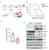
Figure 1
Figure 1
MET overexpression decreases survival of mice harboring EGFR-driven lung adenocarcinoma. (A) Cartoon depicting the strategy to generate EGFR/MET mice (top) and to activate the lung-specific expression of the two transgenes upon exposure to doxycycline diet (bottom). (B) Kaplan–Meier plot showing the survival rate of EGFR (n = 14) and EGFR/MET (n = 12) mice after doxycycline induction; **** p ≤ 0.0001 (Mantel–Cox test). (C) Percentage of adenocarcinomas vs. adenomas in each genotype. Values correspond to the mean ± SEM; * p ≤ 0.05 (unpaired t-test). EGFR (n = 6) and EGFR/MET (n = 5). (D) Immunoblotting of the indicated proteins in lung tumors from EGFR and EGFR/MET mice.
Figure 2
Figure 2
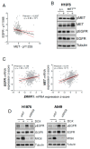
MET expression decreases EGFR activity. (A) Correlation analyses of pMET and pEGFR expression status (Z-scores) in 360 patients from the Lung Adenocarcinoma (TCGA, PanCancer Atlas) dataset. Data were analyzed by Pearson coefficient analysis (r = −0.2037). Linear regression (black line) and 95% confidence intervals (dashed red lines) are also indicated. (B) Immunoblotting of the indicated proteins in H1975 cells infected with lentiviral particles harboring the doxycycline-inducible MET construct (METOX) or empty vector (e.v.). Cells were incubated (+) or not (−) with 1 μg/mL of doxycycline for 48 h. This is a representative example of an experiment performed twice. (C) Correlation analyses for EGFR vs. ERRFI1 (MIG6 gene) and MET vs. ERRFI1 (MIG6 gene) mRNA expression (Z-scores) in 510 patients from the Lung Adenocarcinoma (TCGA, PanCancer Atlas) dataset. Z-scores were analyzed by Pearson coefficient analysis (r = 0.190 and r = 0.314, respectively). Linear regression (black line) and 95% confidence intervals (dashed red lines) are also indicated. (D) Immunoblotting of the indicated proteins in H1975 and A549 cells infected with lentiviral particles harboring the doxycycline-inducible MET construct (METOX) and transfected with non-targeting siRNA (siNT) or siRNA targeting MIG6 (siMIG6). Cells were incubated (+) or not (−) with 1 μg/mL of doxycycline for 48 h. This is a representative example of an experiment performed twice.
Figure 3
Figure 3
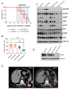
EGFR/MET-driven tumors are resistant to osimertinib. (A) Hematoxylin and eosin staining of whole lungs from EGFR mice treated with vehicle (n = 5) or osimertinib (n = 4) and EGFR/MET mice treated with vehicle (n = 5) or osimertinib (n = 5) for 4 weeks. Scale bar, 5 mm. (B) Tumor burden from same samples as described in (A). Values are the mean ± SEM; *** p ≤ 0.001, **** p ≤ 0.0001 (one-way ANOVA followed by Tukey post hoc test). (C) Immunoblotting of the indicated proteins in lung tumors from EGFR and EGFR/MET mice treated with vehicle or osimertinib. (D) Immunoblotting of the indicated proteins in lungs tumors from EGFR and EGFR/MET mice treated as in (C).
Figure 4
Figure 4
EGFR/MET-driven tumors are sensitive to the osimertinib and crizotinib combination. (A) Kaplan–Meier plot showing the survival rate of EGFR/MET mice treated with vehicle, crizotinib and/or osimertinib (n = 4 mice/group) for 6 weeks. The x axis shows the time since tumor induction by doxycycline; * p ≤ 0.05 (Mantel–Cox test) (B) Tumor burden from the mice described in (A) (n = 4 animals per treatment group). Data are the mean ± SEM; ** p ≤ 0.01 (one-way ANOVA followed by Tukey post hoc test). (C) Immunoblotting of the indicated proteins in lung protein extracts from the same mice described in (A). (D) Immunoblotting of the indicated proteins in lung protein extracts from the same mice described in (A). (E) CT scan of a patient presenting EGFR del19 mutation and MET amplification upon relapse after osimertinib single treatment (CT1) and 3 months after osimertinib and crizotinib treatment started (CT2). Magnification of the tumoral area is presented and highlighted in red.

References

    1. Tan C.S., Gilligan D., Pacey S. Treatment approaches for EGFR-inhibitor-resistant patients with non-small-cell lung cancer. Lancet Oncol. 2015;16:e447–e459. doi: 10.1016/S1470-2045(15)00246-6. - DOI - PubMed
    1. Mok T.S., Wu Y.L., Ahn M.J., Garassino M.C., Kim H.R., Ramalingam S.S., Shepherd F.A., He Y., Akamatsu H., Theelen W.S., et al. Osimertinib or Platinum-Pemetrexed in EGFR T790M-Positive Lung Cancer. N. Engl. J. Med. 2017;376:629–640. doi: 10.1056/NEJMoa1612674. - DOI - PMC - PubMed
    1. Soria J.C., Ohe Y., Vansteenkiste J., Reungwetwattana T., Chewaskulyong B., Lee K.H., Dechaphunkul A., Imamura F., Nogami N., Kurata T., et al. Osimertinib in Untreated EGFR-Mutated Advanced Non-Small-Cell Lung Cancer. N. Engl. J. Med. 2018;378:113–125. doi: 10.1056/NEJMoa1713137. - DOI - PubMed
    1. Piper-Vallillo A.J., Sequist L.V., Piotrowska Z. Emerging Treatment Paradigms for EGFR-Mutant Lung Cancers Progressing on Osimertinib: A Review. J. Clin. Oncol. 2020 doi: 10.1200/JCO.19.03123. - DOI - PubMed
    1. Sequist L.V., Han J.Y., Ahn M.J., Cho B.C., Yu H., Kim S.W., Yang J.C., Lee J.S., Su W.C., Kowalski D., et al. Osimertinib plus savolitinib in patients with EGFR mutation-positive, MET-amplified, non-small-cell lung cancer after progression on EGFR tyrosine kinase inhibitors: Interim results from a multicentre, open-label, phase 1b study. Lancet Oncol. 2020;21:373–386. doi: 10.1016/S1470-2045(19)30785-5. - DOI - PubMed