. 2021 Jul 9;13(14):3442.
doi: 10.3390/cancers13143442.

Interleukin-33-Enhanced CXCR4 Signaling Circuit Mediated by Carcinoma-Associated Fibroblasts Promotes Invasiveness of Head and Neck Cancer

Affiliations

Interleukin-33-Enhanced CXCR4 Signaling Circuit Mediated by Carcinoma-Associated Fibroblasts Promotes Invasiveness of Head and Neck Cancer

Yu-Chun Lin et al. Cancers (Basel). .

Abstract

Despite recent advances, treatment for head and neck squamous cell carcinoma (HNSCC) has limited efficacy in preventing tumor progression. We confirmed previously that carcinoma-associated fibroblasts (CAF)-induced interleukin-33 (IL-33) contributed to cancer progression. However, the molecular mechanisms underlying the complex communication network of the tumor microenvironment merited further evaluation. To simulate the IL-33-induced autocrine signaling, stable clones of IL-33-overexpressing HNSCC cells were established. Besides well-established IL-33/ST2 and SDF1/CXCR4 (stromal-derived factor 1/C-X-C motif chemokine receptor 4) signaling, the CAF-induced IL-33 upregulated CXCR4 via cancer cell induction of IL-33 self-production. The IL-33-enhanced-CXCR4 regulatory circuit involves SDF1/CXCR4 signaling activation and modulates tumor behavior. An in vivo study confirmed the functional role of IL-33/CXCR4 in tumor initiation and metastasis. The CXCR4 and/or IL-33 blockade reduced HNSCC cell aggressiveness, with attenuated invasions and metastases. Immunohistochemistry confirmed that IL-33 and CXCR4 expression correlated significantly with disease-free survival and IL-33-CXCR4 co-expression predicted a poor outcome. Besides paracrine signaling, the CAF-induced IL-33 reciprocally enhanced the autocrine cancer-cell self-production of IL-33 and the corresponding CXCR4 upregulation, leading to the activation of SDF1/CXCR4 signaling subsequent to cancer progression. Thus, targeting the IL-33-enhanced-CXCR4 regulatory circuit attenuates tumor aggressiveness and provides a potential therapeutic option for improving the prognosis in HNSCC patients.

Keywords: carcinoma-associated fibroblast; head and neck squamous cell carcinoma; interleukin-33/CXCR4 regulatory circuit; tumor microenvironment; tumor progression.


Conflict of interest statement

The authors declare no conflict of interest.

Figures

Figure 1
Figure 1
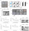
Comparative analysis and characterization of CAFs and HGFs and their effects on two stable clones of the IL-33-overexpressing HNSCC cells and control HNSCC cells. (a) Besides the upregulation of IL-33, ST2, and CXCR4, the expressions of two representative invasion biomarkers, MMP2 and MMP9 by RT-PCR, were enhanced statically, in the cloned IL-33-overexpressing HNSCC cells compared to in the control HNSCC cells. (b) Western blotting analysis showed higher expressions of ST2 and CXCR4 in the cloned IL-33-overexpressing HNSCC cells than in the control HNSCC cells. The uncropped Western blots have been shown in Figure S2. (c) Flow cytometry revealed higher expressions of CXCR4 in the cloned IL-33-overexpressing HNSCC cells than in the control HNSCC cells. (d) The top panel shows the phenotypic differences between the CAFs and HGFs. The Western blotting analysis showed that the expressions of SDF-1 and IL33 in two representative CAFs were statistically higher than that in HGFs (p < 0.05) with further validation by ELISA shown at the bottom panels. (e) Flow cytometry demonstrated that the CAFs displayed higher expressions of CD10 and GPR77 than the HGFs. (f) Four test groups and one control group were classified for a comparative analysis of invasive ability via organotypic culture. The induction of invasion (shown by arrows) demonstrated that there were many foci of the infiltrated cells in the IL-33-overexpressing stable clones, which were markedly enhanced by the CAF-induced IL-33 (in upper and lower center panels). However, in the control group and one test group wherein the IL-33-overexpressing stable clones were cultured without the CAF-induced IL-33, no obvious invasion induction or decreased invasion induction was seen (left two upper and lower panels). The blockade of the IL-33 via shIL-33 or by using an inhibitor of CXCR4 (25 µg/mL AMD3100) markedly decreased the invasion ability (right upper and lower two panels). The statistical analysis of tumor invasion depth is shown in the right panel. * p < 0.05; ** p < 0.01.
Figure 2
Figure 2
Comparative analysis of invasion-related biomarkers associated with the expression of IL-33/CXCR4 signaling between two stable clones of the IL-33-overexpressing HNSCC cells and control. (a) Western blotting analysis demonstrated that increased expressions of IL-33 and CXCR4 were seen in both the IL-33-overexpressing stable clones. Inhibition by shIL-33 markedly decreased the expression of CXCR4; however, the shutdown of CXCR4 did not decrease the expression of IL-33, suggesting that IL-33 expression was mediated upstream of CXCR4 in the signaling cascade. Increased expressions of invasion-related MMP2 and MMP9 were seen in the IL-33-overexpressing stable clones; conversely, shutdown of IL-33 markedly decreased the expression of MMP2 and MMP9. Increased migration (b) and invasion (c) abilities were seen in the cloned IL-33-overexpressing HNSCC cells compared with those of the controls. Blockades with IL-33 and CXCR4 elicited the opposite results. (d) Western blotting analysis showed that blockade of the IL-33 expression decreased the molecules involved in the p38MAPK and pNFκB signaling cascade. In contrast, the inhibition of CXCR4 did not induce similar effects, again suggesting that IL-33 was situated upstream of CXCR4 in the signaling cascade. (e) However, the inhibition of phosphorylated p38 through ERK1/2 and JNK did not affect IL-33 expression, but it decreased CXCR4 expression. * p < 0.05.
Figure 3
Figure 3
Characterization of the two stably cloned IL-33-overexpressing HNSCC cells facilitated by an additional IL-33 expression that led to the induction of the CSC properties. ((a), left) A more aggressive phenotype was seen in the stable clones of the IL-33-overexpressing HNSCC cells (arrows) acting in an autocrine manner, compared with the parent cells. Using the recombinant IL-33 for the paracrine effect of the CAF-induced IL-33, showed a similar effect. There were significant differences in both the stably cloned HNSCC cells and HNSCC cells exposed to the recombinant IL-33 compared with the control HNSCC cells (p < 0.05). ((a), middle and right) Flow cytometric analysis of ALDH1 showed significantly increased activity in both the stably cloned HNSCC cells and HNSCC cells exposed to the recombinant IL-33 compared with the control HNSCC cells (p < 0.05). (b) Western blotting analysis showed the overexpression of CSC-representative markers, including CD44, CD133, Oct-4, and Nanog, in both the stably cloned HNSCC cells and HNSCC cells exposed to the recombinant IL-33 compared with the control HNSCC cells. (c) In the nonadherent sphere culture system, both the stably cloned HNSCC cells and the HNSCC cells exposed to the recombinant IL-33 gradually generated well-formed spheres within seven days—an important property of CSC. However, the control cells yielded only discrete, small cell masses without sphere formation. (d) Chemoresistance was prominent in both the stably cloned HNSCC cells and the HNSCC cells exposed to recombinant IL-33 compared with the control HNSCC cells. Chemosensitivity did not differ significantly between both the stably cloned HNSCC cell lines and the HNSCC cells exposed to recombinant IL-33 compared with the control HNSCC cells (p < 0.05). Western blotting analysis demonstrated that both the stably cloned HNSCC cell lines and the HNSCC cells exposed to the recombinant IL-33 showed an upregulation of the drug resistance-related genes, ABCG-2 and MDR-1. (e) Radioresistance was more prominent in both the stably cloned HNSCC cells and the HNSCC cells exposed to the recombinant IL-33 than in the control HNSCC cells. Radiosensitivity differed significantly between both the stably cloned HNSCC cell lines and the HNSCC cells exposed to the recombinant IL-33 compared with the control HNSCC cells (p < 0.05). Western blotting analysis demonstrated that both the stably cloned HNSCC cell lines and the HNSCC cells exposed to the recombinant IL-33 showed increased antiapoptotic activity, as shown by the upregulation of Bcl-2 and the downregulation of Bax and caspase 3 compared with the HNSCC cells. * Significant difference (p < 0.05) between the experimental and control groups; results are expressed as mean ± SD.
Figure 4
Figure 4
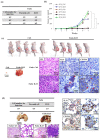
Xerographic evidence from the stable clones of the IL-33-overexpressing HNSCC cells highlights that tumorigenicity and metastases is associated with IL-33 expression/CXCR4 signaling. (a) The in vivo study (n = 5 in each group) showed greater tumorigenicity in the IL-33-overexpressing stably cloned FaDu cells compared with the control cells. (b) Time-dependent tumor growth differences were observed in the stably cloned FaDu cells compared with the control cells. (c) The stably cloned cell-induced tumors (right) were significantly larger than the tumors induced by the control cells (left). A marked size difference in the tumors dissected from a representative BALB/c nude mouse was found following the subcutaneous inoculation of the stably cloned FaDu cells and the control cells. The immunohistochemical analysis of the tumor nodules revealed the overexpression of both IL-33 and CXCR4 in the stably cloned FaDu cells (magnification, ×200). (d) The in vivo study (n = 5 in each group) showed a greater capability for metastasis (3/5) in the IL-33-overexpressing stably cloned FaDu cells than in the control cells (0/5). The gross appearance of the lung tissue dissected from a representative BALB/c nude mouse showed foci of hemorrhagic nodules (arrowheads) in the stable clone cell-induced tumors (right) and no metastatic nodules in the control lung tissue (left). The microscopic findings of the small metastatic hemorrhagic nodules (arrows) in the lung tissue revealed aggregates of many abnormal HNSCC cells with a high nucleus/cytoplasm ratio and a hyperchromatism of the nuclei in the lung parenchyma. Further immunohistochemical analysis of a metastatic tumor nodule in the lung tissue which confirmed their metastatic origin from the HNSCC cells and the overexpression of IL-33 and CXCR4 in the tumor nodules (magnification, ×100 and ×200). In addition, the metastatic small nodules were found in both lungs, and there was no metastases-related involvement of any other organ on necropsy. * p < 0.05.
Figure 5
Figure 5
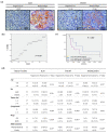
Immunohistochemical analysis of IL-33 and CXCR4 expression and the clinical implications of the IL-33/CXCR4 signaling circuit in 40 patients with HNSCC. (a) Photomicrographs of immunohistochemical staining IL-33 and CXCR4 expression in the HNSCC patients. Twenty-two patients (55%) showed negative or low expression of IL-33, 18 patients (45%) showed a positive immunoreactivity of IL-33 expression, 13 patients (32.5%) showed negative or low expression of CXCR4, and 27 patients (67.5%) showed positive immunoreactivity of CXCR4 (magnification, ×200). (b) The linear regression analysis of IL-33 and CXCR4 expression showed a significant correlation of IL-33 expression with CXCR4 expression (p = 0.027). (c) The Kaplan–Meier analysis of disease-free survival of HNSCC patients after a 60-month follow-up plotted as patients who showed negative immunoreactivity versus positive immunoreactivity for IL-33 and CXCR4 expression in tumor cells. Patients negative for IL-33 and CXCR4 expression had a longer disease-free survival compared with those who tested negative for IL-33 and positive for CXCR4. The co-expression of IL-33 and CXCR4 in HNSCC cells showed a significant correlation with shorter disease-free survival (p < 0.001) which indicated a worse outcome. (d) Moreover, the individual expression of IL-33 and CXCR4 as well as the co-expression of both IL-33 and CXCR4 showed a significant correlation with the clinical parameters including tumor differentiation, lymph node involvement, and TNM stage. * p < 0.05.

References

    1. Bray F., Ferlay J., Soerjomataram I., Siegel R.L., Torre L.A., Jemal A. Global cancer statistics 2018: GLOBOCAN estimates of incidence and mortality worldwide for 36 cancers in 185 countries. CA Cancer J. Clin. 2018;68:394–424. doi: 10.3322/caac.21492. - DOI - PubMed
    1. Karnov K.K.S., Grønhøj C., Jensen D.H., Wessel I., Charabi B.W., Specht L., Kjaer A., von Buchwald C. Increasing incidence and survival in oral cancer: A nationwide Danish study from 1980 to 2014. Acta Oncol. 2017;56:1204–1209. doi: 10.1080/0284186X.2017.1307516. - DOI - PubMed
    1. Kakinuma T., Hwang S.T. Chemokines, Chemokine Receptors, and Cancer Metastasis. J. Leukoc. Biol. 2006;79:639–651. doi: 10.1189/jlb.1105633. - DOI - PubMed
    1. Ben-Baruch A. Site-Specific Metastasis Formation: Chemokines as Regulators of Tumor Cell Adhesion, Motility and Invasion. Cell. Adh Migr. 2009;3:328–333. doi: 10.4161/cam.3.4.9211. - DOI - PMC - PubMed
    1. Teicher B.A., Fricker S.P. CXCL12 (SDF-1)/CXCR4 Pathway in Cancer. Clin. Cancer Res. 2010;16:2927–2931. doi: 10.1158/1078-0432.CCR-09-2329. - DOI - PubMed

LinkOut - more resources