. 2021 Jul 10;13(14):3457.
doi: 10.3390/cancers13143457.

NPM1 Mutational Status Underlines Different Biological Features in Pediatric AML

Affiliations

NPM1 Mutational Status Underlines Different Biological Features in Pediatric AML

Claudia Tregnago et al. Cancers (Basel). .

Abstract

Nucleophosmin (NPM1) is a nucleocytoplasmic shuttling protein, predominantly located in the nucleolus, that regulates a multiplicity of different biological processes. NPM1 localization in the cell is finely tuned by specific signal motifs, with two tryptophan residues (Trp) being essential for the nucleolar localization. In acute myeloid leukemia (AML), several NPM1 mutations have been reported, all resulting in cytoplasmic delocalization, but the putative biological and clinical significance of different variants are still debated. We explored HOXA and HOXB gene expression profile in AML patients and found a differential expression between NPM1 mutations inducing the loss of two (A-like) Trp residues and those determining the loss of one Trp residue (non-A-like). We thus expressed NPM1 A-like- or non-A-like-mutated vectors in AML cell lines finding that NPM1 partially remained in the nucleolus in the non-A-like NPM1-mutated cells. As a result, only in A-like-mutated cells we detected HOXA5, HOXA10, and HOXB5 hyper-expression and p14ARF/p21/p53 pathway deregulation, leading to reduced sensitivity to the treatment with either chemotherapy or Venetoclax, as compared to non-A-like cells. Overall, we identified that the NPM1 mutational status mediates crucial biological characteristics of AML cells, providing the basis for further sub-classification and, potentially, management of this subgroup of patients.

Keywords: HOX genes; Nucleophosmin, NPM1; TP53; acute myeloid leukemia; drug treatment; gene expression; genetic; mutation.


Conflict of interest statement

The authors declare no conflict of interest.

Figures

Figure 1
Figure 1
Intracellular localization of mutated NPM1. (A) Immunofluorescence of HL-60 AML cells transfected with pEGFP-NPM1wt, pEGFP-NPM1mutA, pEGFP-NPM1mutB, pEGFP-NPM1mutD, pEGFP-NPM1mut5, and pEGFP-NPM1mut7 plasmids, where EFGP is fused to NPM1. EGFP signal (green) represents NPM1 protein, and nuclei are stained with DAPI (blue) (40× magnification, scale bar = 15 μm). (B) Western blot showing EGFP protein on nuclear fraction of EGFP positive HL-60 cells transfected with pEGFP-NPM1wt, pEGFP-NPM1mutA, pEGFP-NPM1mutB, pEGFP-NPM1mutD, pEGFP-NPM1mut5, and pEGFP-NPM1mut7 plasmids. The whole western blot images are in Figure S5.
Figure 2
Figure 2
HOXA and HOXB gene expression according to NPM1 mutations. (A) Hierarchical clustering analyses of 71 AML patients for HOXA and HOXB family genes expression. AML genetic subtypes are reported in the legend with color codes. (B) Heatmap visualization of the median expression of HOXA and HOXB genes within the different AML genetic subtypes: NPM1 mutations n = 4, MLL translocations n = 7, NUP98 translocations n = 19, negative for tested molecular markers n = 21, CBF rearrangements n = 20. (C) mRNA expression levels of HOXA5, HOXA10 and HOXB5 in HL-60 cells transfected with pEGFP-NPM1wt, pEGFP-NPM1mutB (A-like), and pEGFP-NPM1mut7 (non-A-like), evaluated by RQ-PCR. Data are presented as mean of three independent experiments ± SEM. * p-value < 0.05, ** p-value < 0.01, *** p-value < 0.001.
Figure 3
Figure 3
Impact of different NPM1 mutations on p53 pathway. (A,B) Western blot showing p14ARF, p21, and p53 protein levels in HL-60 (A) and SHI-1 cells (B) transfected with pEGFP-NPM1wt, pEGFP-NPM1mutB (A-like), and pEGFP-NPM1mut7 (non-A-like) plasmids. GAPDH was used as calibrator. Histograms show protein quantification using ImageJ software, and normalized with respect to GAPDH levels.
Figure 4
Figure 4
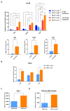
Treatment sensitivity in different NPM1-mutated cells. (A) Percentage of Annexin-V/7AAD positive cells, relative to DMSO, 48 h after Eto (1 µM), AraC (2.5 µM), Ven (5 nM), in HL-60 cells transfected with pEGFP-NPM1mutA, mutB, mutD (A-like) or pEGFP-NPM1mut5, mut7 (non-A-like) plasmids, gating on EGFP positive cells. In the lower panel is reported the mean of Annexin-V/7AAD positive cells after treatment, considering A-like and non-A-like mutations together. * p-value < 0.05, ** p-value < 0.01, *** p-value < 0.001, **** p-value < 0.0001. (B) Percentage of Annexin-V/7AAD positive cells in SHI-1 transfected with pEGFP-NPM1mutB (A-like) or pEGFP-NPM1mut7 (non-A-like) plasmids, gating on EGFP positive cells and relative to DMSO, after 48-h of treatment with Eto (1 µM), AraC (2.5 µM), or Ven (1 µM). * p-value < 0.05. (C) Reduction of cell viability measured by ATP assay after 24 h of treatment with Eto (1 µM), relative to DMSO, in SHI-1 cells transfected with pEGFP-NPM1mutB (A-like) or pEGFP-NPM1mut7 (non-A-like) vectors. * p-value < 0.05. (D) Reduction of cell viability measured by ATP assay after 24-h treatment with AraC (2.5 µM), relative to DMSO, in primary AML samples harboring A-like or non-A-like NPM1 mutations. ** p-value < 0.01. rlu: relative luminescence unit.

References

    1. Cordell J.L., Pulford K.A.F., Bigerna B., Roncador G., Banham A., Colombo E., Pelicci P.G., Mason D.Y., Falini B. Detection of normal and chimeric nucleophosmin in human cells. Blood. 1999;93:632–642. doi: 10.1182/blood.V93.2.632. - DOI - PubMed
    1. Lam Y.W., Trinkle-Mulcahy L., Lamond A.I. The nucleolus. J. Cell Sci. 2005;118:1335–1337. doi: 10.1242/jcs.01736. - DOI - PubMed
    1. Poletto M., Lirussi L., Wilson D.M., Tell G. Nucleophosmin modulates stability, activity, and nucleolar accumulation of base excision repair proteins. Mol. Biol. Cell. 2014;25:1641–1652. doi: 10.1091/mbc.e13-12-0717. - DOI - PMC - PubMed
    1. Vascotto C., Lirussi L., Poletto M., Tiribelli M., Damiani D., Fabbro D., Damante G., Demple B., Colombo E., Tell G. Functional regulation of the apurinic/apyrimidinic endonuclease 1 by nucleophosmin: Impact on tumor biology. Oncogene. 2014;33:2876–2887. doi: 10.1038/onc.2013.251. - DOI - PubMed
    1. Box J.K., Paquet N., Adams M.N., Boucher D., Bolderson E., O’Byrne K.J., Richard D.J. Nucleophosmin: From structure and function to disease development. BMC Mol. Biol. 2016;17:19. doi: 10.1186/s12867-016-0073-9. - DOI - PMC - PubMed