. 2021 Jul 7;22(14):7290.
doi: 10.3390/ijms22147290.

Chondroprotective Effects of a Histone Deacetylase Inhibitor, Panobinostat, on Pain Behavior and Cartilage Degradation in Anterior Cruciate Ligament Transection-Induced Experimental Osteoarthritic Rats

Affiliations

Chondroprotective Effects of a Histone Deacetylase Inhibitor, Panobinostat, on Pain Behavior and Cartilage Degradation in Anterior Cruciate Ligament Transection-Induced Experimental Osteoarthritic Rats

Zhi-Hong Wen et al. Int J Mol Sci. .

Abstract

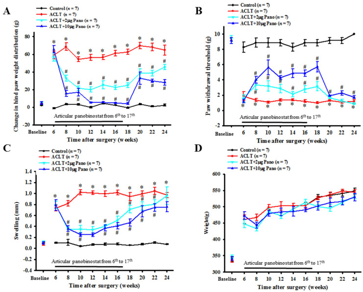
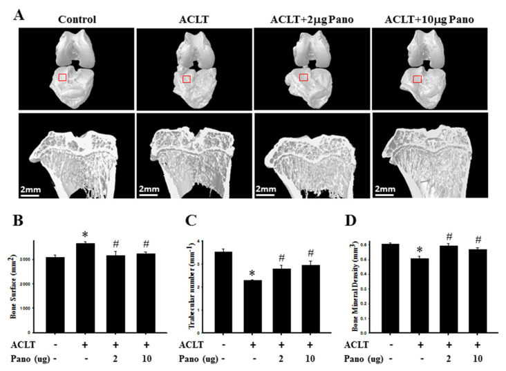
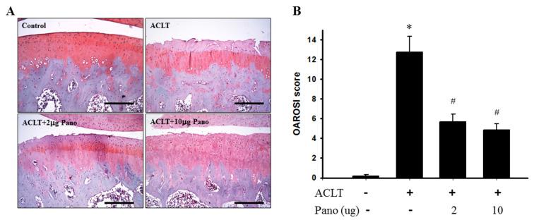
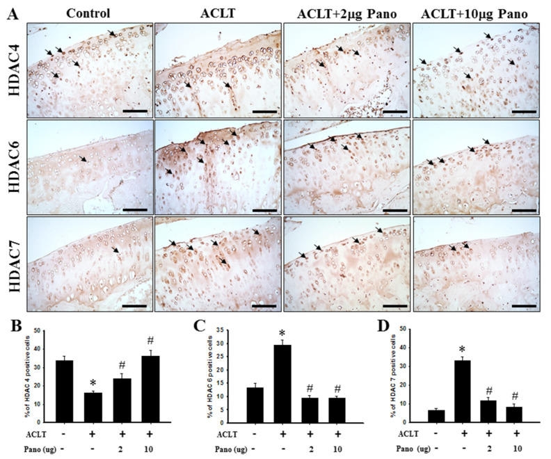
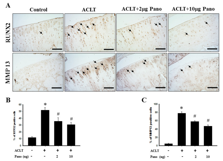
Osteoarthritis (OA) is the most common articular degenerative disease characterized by chronic pain, joint inflammation, and movement limitations, which are significantly influenced by aberrant epigenetic modifications of numerous OA-susceptible genes. Recent studies revealed that both the abnormal activation and differential expression of histone deacetylases (HDACs) might contribute to OA pathogenesis. In this study, we investigated the chondroprotective effects of a marine-derived HDAC inhibitor, panobinostat, on anterior cruciate ligament transection (ACLT)-induced experimental OA rats. The intra-articular administration of 2 or 10 µg of panobinostat (each group, n = 7) per week from the 6th to 17th week attenuates ACLT-induced nociceptive behaviors, including secondary mechanical allodynia and weight-bearing distribution. Histopathological and microcomputed tomography analysis showed that panobinostat significantly prevents cartilage degeneration after ACLT. Moreover, intra-articular panobinostat exerts hypertrophic effects in the chondrocytes of articular cartilage by regulating the protein expressions of HDAC4, HDAC6, HDAC7, runt-domain transcription factor-2, and matrix metalloproteinase-13. The study indicated that HDACs might have different modulations on the chondrocyte phenotype in the early stages of OA development. These results provide new evidence that panobinostat may be a potential therapeutic drug for OA.

Keywords: histone deacetylases; nociception; osteoarthritis; panobinostat.


Conflict of interest statement

The authors declare no conflict of interest.

Figures

Figure 1
Figure 1
Effects of panobinostat on ACLT-induced OA model. Time courses of the effects of panobinostat on (A) ACLT-induced hind paw weight-bearing deficits, (B) mechanical allodynia, (C) knee swelling, and (D) rat weight. The rats in the control group did not receive surgery and treatment, whereas rats in the ACLT group underwent ACLT. Rats in the ACLT + panobinostat groups were intra-articularly injected with panobinostat (2 or 10 µg per week) from the 6th to 17th week after ACLT, whereas the other groups received an intra-articular injection of vehicle. Each value is presented as means ± SEM for each group. ACLT, anterior cruciate ligament transection; OA, osteoarthritis; Pano, panobinostat; SEM, standard error of the mean. (* p < 0.05 vs. the control group; # p < 0.05 vs. the ACLT group).
Figure 2
Figure 2
Micro-CT analysis of the bone structure in ACLT model and panobinostat treatment. (A) Representative three-dimensional renderings of the tibial and femoral condyles (upper panel) and sagittal views (middle panel) scanned via micro-CT. The sagittal view of micro-CT images show changes in osteoarthritic differences in the subchondral bone structures of medial femoral and tibial compartments. Red frame is the region of tibial plateau in the knee joint. (B) Quantitative analysis of bone surface (mm2), (C) trabecular number (mm−1), and (D) bone mineral density (mm3). ACLT, anterior cruciate ligament transection; Pano, panobinostat. (* p < 0.05 vs. the control group; # p < 0.05 vs. the ACLT group).
Figure 3
Figure 3
Histopathological evaluation of the tibia in knee joints after panobinostat treatment in an ACLT rat model. (A) Safranin O/Fast Green staining was performed on the histological sections of knee joints from the control, ACLT, and ACLT + panobinostat (2 or 10 μg) groups. Representative images of Safranin O/Fast Green staining for articular cartilage show cartilage damages in the ACLT knee compared with panobinostat treatment. The scale bar represents 250 μm. (B) Histopathological changes in the knee joints of the four studied groups were evaluated using the OARSI scoring system. Histogram shows the OARSI scores of the control, ACLT, and ACLT + panobinostat (2 or 10 μg) groups. OARSI, Osteoarthritis Research Society International; ACLT, anterior cruciate ligament transection; Pano, panobinostat. (* p < 0.05 vs. the control group; # p < 0.05 vs. the ACLT group).
Figure 4
Figure 4
Effects of panobinostat on the expression of HDAC4, HDAC6, and HDAC7 in cartilage tissues. (A) Immunohistochemical staining of HDAC4, HDAC6, and HDAC7 in joint sections from the control, ACLT, ACLT + panobinostat (2 or 10 μg) groups. The immunoreactive positive cells are indicated in brown (arrows). Results from the quantitative analysis of the ratio of (B) HDAC4-, (C) HDAC6-, and (D) HDAC7-positive cells in joint sections are presented. Data are expressed as means ± SEM for each group. Scale bar represents 100 μm. HDAC, histone deacetylase; ACLT, anterior cruciate ligament transection; Pano, panobinostat; SEM, standard error of the mean. (* p < 0.05 vs. the control group; # p < 0.05 vs. the ACLT group).
Figure 5
Figure 5
Effects of panobinostat on the expression of RUNX2 and MMP13 in cartilage tissues. (A) Immunohistochemical staining of RUNX2 and MMP13 in joint sections from the control, ACLT, ACLT + panobinostat (2 or 10 μg) groups. The immunoreactive positive cells are indicated in brown (arrows). Results from the quantitative analysis of the ratio of (B) RUNX2- and (C) MMP13-positive cells in joint sections are presented. Data are expressed as means ± SEM for each group. Scale bar represents 100 μm. RUNX2, runt-domain transcription factor-2; MMP13, matrix metalloproteinase-13; ACLT, anterior cruciate ligament transection; Pano, panobinostat; SEM, standard error of the mean. (* p < 0.05 vs. the control group; # p < 0.05 vs. the ACLT group).

References

    1. Garnock-Jones K.P. Panobinostat: First global approval. Drugs. 2015;75:695–704. doi: 10.1007/s40265-015-0388-8. - DOI - PubMed
    1. Rubiolo J., Alonso E., Cagide E. Seafood and Freshwater Toxins. CRC Press; Boca Raton, FL, USA: 2014. Marine compounds as a starting point to drugs; pp. 1141–1178.
    1. Conte M., Fontana E., Nebbioso A. Marine-derived secondary metabolites as promising epigenetic bio-compounds for anticancer therapy. Mar. Drugs. 2020;19:15. doi: 10.3390/md19010015. - DOI - PMC - PubMed
    1. Wang Y.Q., Miao Z.H. Marine-derived angiogenesis inhibitors for cancer therapy. Mar. Drugs. 2013;11:903–933. doi: 10.3390/md11030903. - DOI - PMC - PubMed
    1. Quiñoà E., Crews P. Phenolic constituents of Psammaplysilla. Tetrahedron Lett. 1987;28:3229–3232. doi: 10.1016/S0040-4039(00)95478-9. - DOI

MeSH terms

Substances