

Neither a Novel Tau Proteinopathy nor an Expansion of a Phenotype: Reappraising Clinicopathology-Based Nosology
- PMID: 34298918
- PMCID: PMC8329925
- DOI: 10.3390/ijms22147292
Neither a Novel Tau Proteinopathy nor an Expansion of a Phenotype: Reappraising Clinicopathology-Based Nosology
Abstract
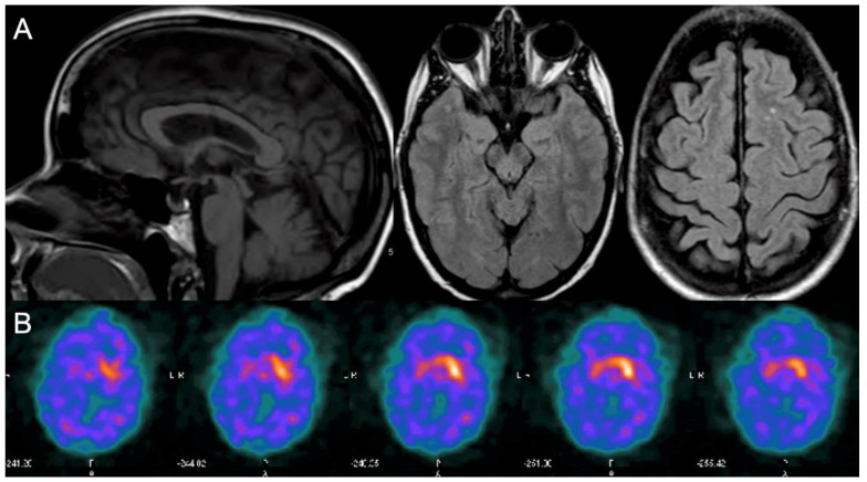
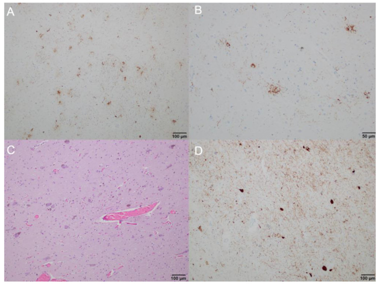
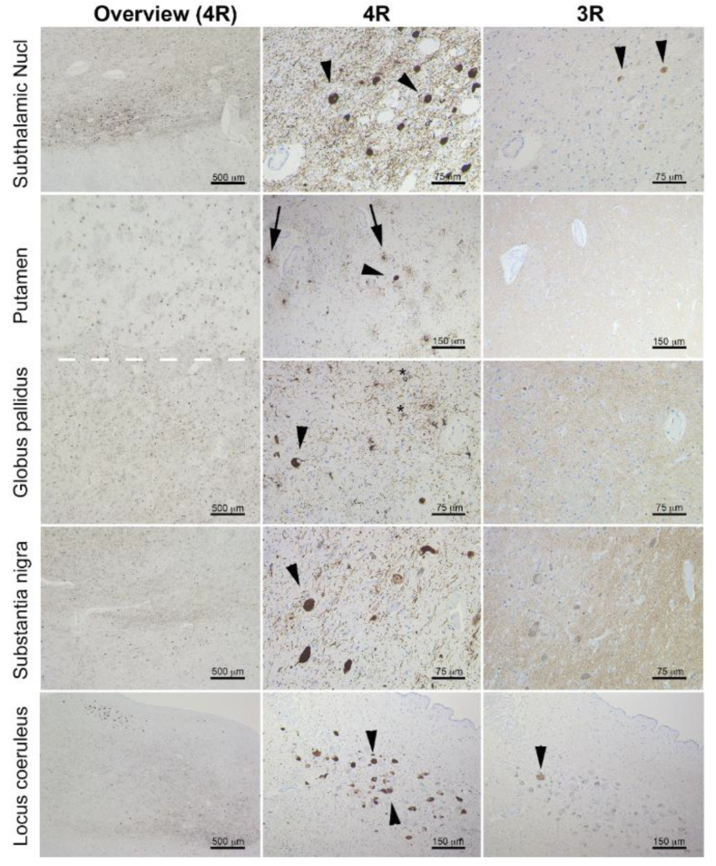
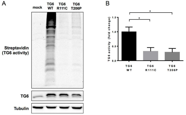
The gold standard for classification of neurodegenerative diseases is postmortem histopathology; however, the diagnostic odyssey of this case challenges such a clinicopathologic model. We evaluated a 60-year-old woman with a 7-year history of a progressive dystonia-ataxia syndrome with supranuclear gaze palsy, suspected to represent Niemann-Pick disease Type C. Postmortem evaluation unexpectedly demonstrated neurodegeneration with 4-repeat tau deposition in a distribution diagnostic of progressive supranuclear palsy (PSP). Whole-exome sequencing revealed a new heterozygous variant in TGM6, associated with spinocerebellar ataxia type 35 (SCA35). This novel TGM6 variant reduced transglutaminase activity in vitro, suggesting it was pathogenic. This case could be interpreted as expanding: (1) the PSP phenotype to include a spinocerebellar variant; (2) SCA35 as a tau proteinopathy; or (3) TGM6 as a novel genetic variant underlying a SCA35 phenotype with PSP pathology. None of these interpretations seem adequate. We instead hypothesize that impairment in the crosslinking of tau by the TGM6-encoded transglutaminase enzyme may compromise tau functionally and structurally, leading to its aggregation in a pattern currently classified as PSP. The lessons from this case study encourage a reassessment of our clinicopathology-based nosology.
Keywords: cerebellar ataxia; movement disorders; neurogenetics; postmortem.
Conflict of interest statement
The authors declare that they have no conflict of interest related to the research covered in this article. Luca Marsili has received honoraria from the International Association of Parkinsonism and Related Disorders (IAPRD) Society for social media and web support. Alberto J Espay has received grant support from the NIH and the Michael J Fox Foundation; personal compensation as a consultant/scientific advisory board member for Abbvie, Neuroderm, Neurocrine, Amneal, Adamas, Acadia, Acorda, Kyowa Kirin, Sunovion, Lundbeck, and USWorldMeds; publishing royalties from Lippincott Williams & Wilkins, Cambridge University Press, and Springer; and honoraria from USWorldMeds, Acadia, and Sunovion. Espay is cofounder of REGAIN Therapeutics, owner of a provisional patent on compositions and methods for treatment and/or prophylaxis of proteinopathies. Christopher D. Stephen has provided scientific advisory for Xenon Pharmaceuticals and SwanBio Therapeutics and received research funding from Sanofi-Genzyme for a study of video oculography in late-onset GM2 gangliosidosis. Gabor G. Kovacs has served as an advisor for Biogen and received publishing royalties from Elsevier, Wiley-Blackwell, and Cambridge University Press, and shares a patent for the a-synculein antibody 5G4. Anthony E. Lang has served as an advisor for Abbvie, Acorda, AFFiRis, Biogen, Denali, Janssen, Lilly, Lundbeck, Maplight, Paladin, Retrophin, Roche, Sun Pharma, Sunovion, Theravance, and Corticobasal Degeneration Solutions; received honoraria from Sun Pharma, AbbVie and Sunovion; received grants from Brain Canada, Canadian Institutes of Health Research, Corticobasal Degeneration Solutions, Edmond J Safra Philanthropic Foundation, Michael J. Fox Foundation, the Ontario Brain Institute, Parkinson Foundation, Parkinson Canada, and W. Garfield Weston Foundation; received publishing royalties from Elsevier, Saunders, Wiley-Blackwell, Johns Hopkins Press, and Cambridge University Press. Marcelo A. Kauffman is an employee of the CONICET and has received grant support from the Ministry of Science and Technology of Argentina and the Ministry of Health of Buenos Aires. Andrea Sturchio is cofounder of REGAIN Therapeutics, owner of a provisional patent on compositions and methods for treatment and/or prophylaxis of proteinopathies.
Figures
References
-
- Song Y., Kirkpatrick L.L., Schilling A.B., Helseth D.L., Chabot N., Keillor J.W., Johnson G.V., Brady S.T. Transglutaminase and polyamination of tubulin: Posttranslational modification for stabilizing axonal microtubules. Neuron. 2013;78:109–123. doi: 10.1016/j.neuron.2013.01.036. - DOI - PMC - PubMed
Publication types
MeSH terms
Substances
Grants and funding
LinkOut - more resources
Full Text Sources
Miscellaneous
