Review
. 2021 Jul 7;22(14):7319.
doi: 10.3390/ijms22147319.

New Insights into Multiple Sclerosis Mechanisms: Lipids on the Track to Control Inflammation and Neurodegeneration

Affiliations
Review

New Insights into Multiple Sclerosis Mechanisms: Lipids on the Track to Control Inflammation and Neurodegeneration

Maria Podbielska et al. Int J Mol Sci. .

Abstract

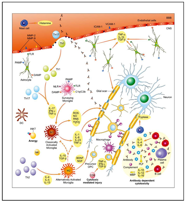
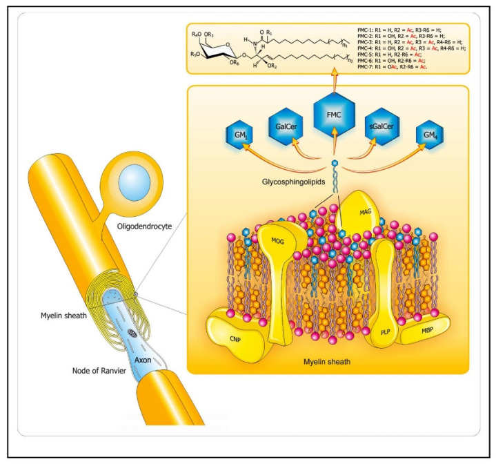
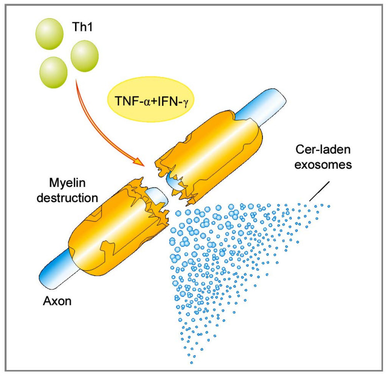
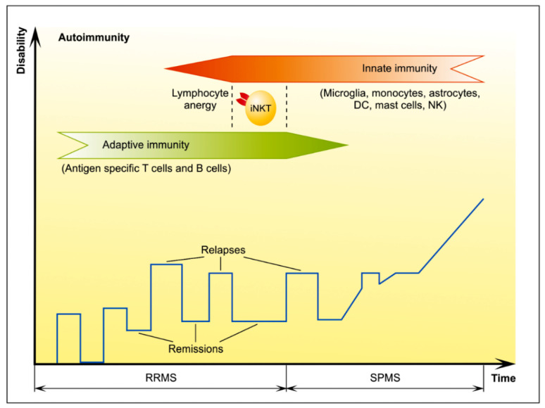
Multiple sclerosis (MS) is a central nervous system disease with complex pathogenesis, including two main processes: immune-mediated inflammatory demyelination and progressive degeneration with axonal loss. Despite recent progress in our understanding and management of MS, availability of sensitive and specific biomarkers for these both processes, as well as neuroprotective therapeutic options targeted at progressive phase of disease, are still being sought. Given their abundance in the myelin sheath, lipids are believed to play a central role in underlying immunopathogenesis in MS and seem to be a promising subject of investigation in this field. On the basis of our previous research and a review of the literature, we discuss the current understanding of lipid-related mechanisms involved in active relapse, remission, and progression of MS. These insights highlight potential usefulness of lipid markers in prediction or monitoring the course of MS, particularly in its progressive stage, still insufficiently addressed. Furthermore, they raise hope for new, effective, and stage-specific treatment options, involving lipids as targets or carriers of therapeutic agents.

Keywords: MS biomarkers; MS mechanisms; MS therapy; central nervous system; inflammation; lipidomics; lipids; multiple sclerosis; neurodegeneration; neurological diseases.


Conflict of interest statement

The authors declare no conflict of interest.

Figures

Figure 1
Figure 1
The core of multiple sclerosis (MS) background is associated with disturbed, autoreactive activity of both the innate and adaptive immunological system. As a result of complex interplay between genetic and environmental factors, pools of auto-reactive T cells are activated and enter the central nervous system (CNS) through the disrupted blood–brain barrier (BBB). Their entry is facilitated, i.e., by enhanced expression of endothelial adhesion molecules (ICAM-1, VCAM-1) and matrix metalloproteinases (MMP-2, MMP-9). An activation of glial cells further contributes to pro-inflammatory properties of the CNS environment. Multiple mechanisms of immune-mediated injury of myelin and axons have been postulated: cytokine-mediated damage, digestion of surface myelin antigens by macrophages, antibody-dependent and complement-mediated cytotoxicity, and direct cytotoxic attack by CD8+ T cells. Parallel to inflammatory activity, there is slowly expanding neurodegenerative injury with axonopathy. The main contributing factors include: toxic metabolites (ROS, NO, RNS), mitochondrial and peroxysomal dysfunction with energetic deficit as well as disturbed ionic balance, and emerging pro-apoptotic activity. Abbreviations: BDNF—brain-derived neurotrophic factor, DAMP—danger associated molecular pattern, DC—dendritic cell, IFN-γ—interferon γ, IL—interleukin, iNKT cells—invariant natural killer T cells, MBP—myelin basic protein, NGF—nerve growth factor, NLR—NOD-like receptors, PAMP—pathogen-associated molecular pattern, TGF-β—transforming growth factor β, Th—T helper, TLR—Toll-like receptor, TNF-α—tumor necrosis factor α. Adapted from [5].
Figure 2
Figure 2
Lipid distribution in the CNS myelin sheaths. The diagram depicts arrangement of complex lipids (cholesterol, phospholipids, and glycosphingolipids) within the most abundant proteins (PLP, MBP) in CNS myelin. The relative molar constancy of lipids: cholesterol/phospholipids (PLs)/galactosylceramide (GalCer) is 2:2:1. Proteins are marked in yellow, and the comprising lipids are as follows: cholesterol in orange, PLs in pink, and the glycosphingolipids (FMC, fast migrating cerebrosides; GalCer, galactosylceramide; GM1, mono-sialoganglioside; GM4, sialosyl-galactosylceramide; sGalCer, sulfatide) in blue. Structures of unique sphingosine 3-O-acetylated-GalCer glycolipid series, namely, acetyl-cerebrosides (FMCs) are shown at the top. Abbreviations: CNP—2′3′-cyclic-nucleotide 3′-phospodiesterase, MAG—myelin-associated glycoprotein, MBP—myelin basic protein, MOG—myelin oligodendrocyte glycoprotein, PLP—proteolipid protein. Adapted from [5].
Figure 3
Figure 3
Cer-laden exosomes drive acute inflammatory demyelination in MS. Abbreviations: Cer—ceramide, IFN-γ—interferon γ, MS—multiple sclerosis, Th—T helper, TNF-α—tumor necrosis factor α. Adapted from [36].
Figure 4
Figure 4
The iNKT cells act as a bridge of adaptive and innate immunity. A clinical hallmark of MS is the heterogeneous presentation ranging from benign with little or no disability even years after disease onset, a commonly encountered a relapsing-remitting (RR) phase followed by a secondary progressive (SP) phase, progressively disabling course. The RR phase is driven by the adaptive immune response while SP phase is driven by the innate immune system. We propose invariant natural killer T (iNKT) cells act as a bridge between adaptive and innate immunity, the phase characterized by lymphocyte anergy resulting in MS remission. Abbreviations: DC—dendritic cell, NK—natural killer, iNKT cells—invariant natural killer T cells, RRMS—relapsing–remitting multiple sclerosis, SPMS—secondary progressive multiple sclerosis. Adapted and modified from [5].

References

    1. Lublin F.D., Reingold S.C., Cohen J.A., Cutter G.R., Sorensen P.S., Thompson A.J., Wolinsky J.S., Balcer L.J., Banwell B., Barkhof F., et al. Defining the clinical course of multiple sclerosis: The 2013 revisions. Neurology. 2014;83:278–286. doi: 10.1212/WNL.0000000000000560. - DOI - PMC - PubMed
    1. Lassmann H. New concepts on progressive multiple sclerosis. Curr. Neurol. Neurosci. Rep. 2007;7:239–244. doi: 10.1007/s11910-007-0036-0. - DOI - PubMed
    1. Trapp B.D., Nave K.A. Multiple sclerosis: An immune or neurodegenerative disorder? Annu. Rev. Neurosci. 2008;31:247–269. doi: 10.1146/annurev.neuro.30.051606.094313. - DOI - PubMed
    1. Lassmann H. Pathogenic mechanisms associated with different clinical courses of multiple sclerosis. Front. Immunol. 2018;9:3116. doi: 10.3389/fimmu.2018.03116. - DOI - PMC - PubMed
    1. Podbielska M., O’Keeffe J., Hogan E.L. Autoimmunity in multiple sclerosis: Role of sphingolipids, invariant NKT cells and other immune elements in control of inflammation and neurodegeneration. J. Neurol. Sci. 2018;385:198–214. doi: 10.1016/j.jns.2017.12.022. - DOI - PubMed

MeSH terms