Comparative Study
. 2021 Jul 8;22(14):7368.
doi: 10.3390/ijms22147368.

Biocompatibility and Biological Performance Evaluation of Additive-Manufactured Bioabsorbable Iron-Based Porous Suture Anchor in a Rabbit Model

Affiliations
Comparative Study

Biocompatibility and Biological Performance Evaluation of Additive-Manufactured Bioabsorbable Iron-Based Porous Suture Anchor in a Rabbit Model

Chien-Cheng Tai et al. Int J Mol Sci. .

Abstract

This study evaluated the biocompatibility and biological performance of novel additive-manufactured bioabsorbable iron-based porous suture anchors (iron_SAs). Two types of bioabsorbable iron_SAs, with double- and triple-helical structures (iron_SA_2_helix and iron_SA_3_helix, respectively), were compared with the synthetic polymer-based bioabsorbable suture anchor (polymer_SAs). An in vitro mechanical test, MTT assay, and scanning electron microscope (SEM) analysis were performed. An in vivo animal study was also performed. The three types of suture anchors were randomly implanted in the outer cortex of the lateral femoral condyle. The ultimate in vitro pullout strength of the iron_SA_3_helix group was significantly higher than the iron_SA_2_helix and polymer_SA groups. The MTT assay findings demonstrated no significant cytotoxicity, and the SEM analysis showed cells attachment on implant surface. The ultimate failure load of the iron_SA_3_helix group was significantly higher than that of the polymer_SA group. The micro-CT analysis indicated the iron_SA_3_helix group showed a higher bone volume fraction (BV/TV) after surgery. Moreover, both iron SAs underwent degradation with time. Iron_SAs with triple-helical threads and a porous structure demonstrated better mechanical strength and high biocompatibility after short-term implantation. The combined advantages of the mechanical superiority of the iron metal and the possibility of absorption after implantation make the iron_SA a suitable candidate for further development.

Keywords: additive manufacturing (3D printing); bioabsorbable; iron-based; suture anchor.


Conflict of interest statement

The authors declare no conflict of interest.

Figures
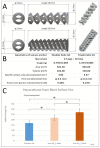
Figure 1
Figure 1
Illustration of iron-based bioabsorbable porous double- and triple-helical suture anchors (A). Geometrical specifications of iron-based bioabsorbable porous double- and triple-helical suture anchors (B). In vitro mechanical ultimate pullout strength test of three types of suture anchors (C). The error bar represents the standard deviation, and * denotes statistical significance between the two groups.
Figure 2
Figure 2
The 3-(4,5-dimethylthiazol-2-yl)-2,5-diphenyl tetrazolium bromide (MTT) assays were performed for the extracts of iron-based bioabsorbable porous suture anchors of different groups (A) which showed >70% cell viability. Scanning electron microscope (SEM) analysis of iron-based bioabsorbable porous testing suture anchors (prototype, not final version) (B) demonstrated cell attachment on the implant surface. Yellow arrows indicated the lamellipodial and filopodial extrusions from the cells (CE). The error bar represented the standard deviation.
Figure 3
Figure 3
In vivo biomechanical ultimate pullout strength assessment for different suture anchors. The ultimate failure load of the iron SA_3 helix group at 4 weeks was significantly higher than those of the iron SA_2 helix and Polymer SA groups. At 12 weeks, the ultimate failure load of the iron SA_2 helix and iron SA_3 helix groups were significantly higher than that of the polymer SA group The error bar represents the standard deviation, and * denotes statistical significance between the two groups.
Figure 4
Figure 4
Micro-computed tomography (Micro-CT) analysis. The region of interest (ROI; diameter, 6.25 mm; thickness, 5 mm) of the bone–implant block was segmented before bone analysis, and bone growth was examined 0–1000 μm around the implant (A). Quantitative evaluation of bone volume between bone and suture anchors. Tissue volume (TV, mm3), bone volume (BV, mm3), and bone surface (BS, mm2) were examined 0–1000 μm above the implant surface. Bone volume fraction (BV/TV) (B) and bone surface density (BS/TV) (C) represent the bone volume rate and bone tissue surface rate, respectively. The error bar represents the standard deviation.
Figure 5
Figure 5
Micro-computed tomography (CT) degradation analysis of the iron suture groups. ST decreased (A) and SSV/OV percentage increased (B) sequentially from the preoperative period until 12 weeks postoperatively. Reconstructed micro-CT images observed preoperatively and at 12 weeks postoperatively (C). ST: structure thickness (mm), SSV: small implant fragments < 0.18 mm in diameter (small-structure volume), OV: object volume, and SSV/OV (%) denoted the small fragment percentage of the implant in the region of interest. The error bar represents the standard deviation.
Figure 6
Figure 6
Blood concentrations of iron (μg/dL), alanine transaminase (ALT; U/L), creatinine (Cr; mg/dL), and blood urea nitrogen (BUN; mg/dL) preoperatively and 1, 2, and 3 months postoperatively. The error bar represented the standard deviation.
Figure 7
Figure 7
Histological examination of the bone–suture anchor interface. The specimens were stained with Sanderson’s rapid bone stain and then counterstained with acid fuchsin. Scale from 12.5×, 40×, and 100×, respectively. B: bone, S: suture anchor.
Figure 8
Figure 8
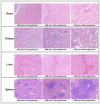
Histopathological examinations of visceral organs (heart, kidney, liver, and spleen). The specimens were obtained from one of the animals implanted with an iron bioabsorbable triple-helix suture anchor. Scale from 40×, 100×, and 400×, respectively. H&E stain: hematoxylin and eosin stain.
Figure 9
Figure 9
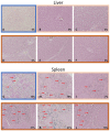
Prussian blue staining of the liver (AF) and spleen (GL). The specimens were obtained from the polymer SA group (A,G), iron SA_2 helix group (B,C,H,I), and iron SA_3 helix group (DF,JL), respectively. Red arrows indicate identified iron stain clusters. Magnification: 200×.

References

    1. Park J.B., Bronzino J.D. Biomaterials: Principles and Applications. 1st ed. CRC Press; Boca Raton, FL, USA: 2002.
    1. Joung Y.-H. Development of implantable medical devices: From an engineering perspective. Int. Neurourol. J. 2013;17:98. doi: 10.5213/inj.2013.17.3.98. - DOI - PMC - PubMed
    1. Khan W., Muntimadugu E., Jaffe M., Domb A.J. Focal Controlled Drug Delivery. Springer; Berlin/Heidelberg, Germany: 2014. Implantable medical devices; pp. 33–59. - DOI
    1. Han H.-S., Loffredo S., Jun I., Edwards J., Kim Y.-C., Seok H.-K., Witte F., Mantovani D., Glyn-Jones S. Current status and outlook on the clinical translation of biodegradable metals. Mater. Today. 2019;23:57–71. doi: 10.1016/j.mattod.2018.05.018. - DOI
    1. Shuai C., Li S., Peng S., Feng P., Lai Y., Gao C. Biodegradable metallic bone implants. Mater. Chem. Front. 2019;3:544–562. doi: 10.1039/C8QM00507A. - DOI

Publication types

MeSH terms

LinkOut - more resources