Review
. 2021 Jul 10;22(14):7425.
doi: 10.3390/ijms22147425.

Structural Evaluation of the Spike Glycoprotein Variants on SARS-CoV-2 Transmission and Immune Evasion

Affiliations
Review

Structural Evaluation of the Spike Glycoprotein Variants on SARS-CoV-2 Transmission and Immune Evasion

Mohd Zulkifli Salleh et al. Int J Mol Sci. .

Abstract

The emergence of severe acute respiratory syndrome coronavirus 2 (SARS-CoV-2) presents significant social, economic and political challenges worldwide. SARS-CoV-2 has caused over 3.5 million deaths since late 2019. Mutations in the spike (S) glycoprotein are of particular concern because it harbours the domain which recognises the angiotensin-converting enzyme 2 (ACE2) receptor and is the target for neutralising antibodies. Mutations in the S protein may induce alterations in the surface spike structures, changing the conformational B-cell epitopes and leading to a potential reduction in vaccine efficacy. Here, we summarise how the more important variants of SARS-CoV-2, which include cluster 5, lineages B.1.1.7 (Alpha variant), B.1.351 (Beta), P.1 (B.1.1.28/Gamma), B.1.427/B.1.429 (Epsilon), B.1.526 (Iota) and B.1.617.2 (Delta) confer mutations in their respective spike proteins which enhance viral fitness by improving binding affinity to the ACE2 receptor and lead to an increase in infectivity and transmission. We further discuss how these spike protein mutations provide resistance against immune responses, either acquired naturally or induced by vaccination. This information will be valuable in guiding the development of vaccines and other therapeutics for protection against the ongoing coronavirus disease 2019 (COVID-19) pandemic.

Keywords: COVID-19; SARS-CoV-2; immune evasion; spike mutations; spike variants; transmission.


Conflict of interest statement

The authors declare no conflict of interest.

Figures
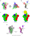
Figure 1
Figure 1
Structures of the severe acute respiratory syndrome coronavirus (SARS-CoV) spike glycoprotein and comparison with the severe acute respiratory syndrome coronavirus 2 (SARS-CoV-2) spike (S) glycoprotein. (a) Side and top views of the inactive prefusion structure of the SARS-CoV S glycoprotein, which adopts a ‘mushroom-like’, homotrimeric arrangement of S1/S2 subunits: chain A (green), chain B (blue) and chain C (red). (b) A single chain of the S glycoprotein which consists of S1 subunit: N-terminal domain (NTD) (red), the receptor-binding domain (RBD) of C-terminal domain CTD1 (blue), CTD2 (green) and CTD3 (yellow), and S2 subunit (black). (c) Three different conformational states of the SARS-CoV S glycoprotein: inactive S homotrimeric glycoprotein complex with all three CTD1 ‘down’ (PDB: 6ACC), active S glycoprotein with one CTD1 ‘up’ (PDB: 6ACD) and the spike-angiotensin-converting enzyme 2 (ACE2) complex (ACE2 shown in yellow) (PDB: 6ACG) [37]. (d) Superimpositions between the SARS-CoV-2 (PDB: 6VSB) [38] and SARS-CoV S glycoproteins. Left panel: SARS-CoV-2 (green), SARS-CoV (magenta). Right panel: chain A of the SARS-CoV-2 S glycoprotein (NTD is in red, CTD1 is in blue, CTD2 is in green, CTD3 is in yellow and S2 is in black) superimposition onto the SARS-CoV S glycoprotein (magenta) using SSM matching, as implemented in CCP4MG; root-mean-square deviation (RMSD): 1.97 Å over 884 Cα atoms.
Figure 2
Figure 2
The SARS-CoV and SARS-CoV-2 RBD/ACE2 complexes. (a) Left: the overall structure of the SARS-CoV RBD (red) bound to ACE2 (yellow). The region contains a receptor-binding motif (RBM) which is highlighted with a box. Middle: L472 of the SARS-CoV RBD interacts with L79 and M82 of ACE2, N479 of the SARS-CoV RBD with H34 of ACE2, a hydrogen bond between G488 of the SARS-CoV RBD and K353 of ACE2. There is no interaction of the SARS-CoV RBD V404 (equivalent to K417 in the SARS-CoV-2 RBD) with any residues from ACE2. Right: the electrostatic potential map of the SARS-CoV RBD, which shows an inward concave structure with the N-terminal helix of ACE2, shown in yellow (PDB: 2AJF) [40]. (b) Left: the overall structure of the SARS-CoV-2 RBD (blue) bound to ACE2 (yellow). Note the similarity between the two RBDs: the core subdomain consists of 5 antiparallel β strands (β1—4 and β7) and is conserved. Middle: F486 of the SARS-CoV-2 RBD interacts with Q24, L79 and Y83 of ACE2, a hydrogen bond between N501 of the SARS-CoV-2 RBD and Y41 of ACE2, two hydrogen bonds between Q493 of the SARS-CoV-2 RBD and E35 of ACE2, a salt-bridge between K417 of the SARS-CoV-2 RBD and D30 of ACE2. Right: the electrostatic potential map of the SARS-CoV-2 RBD with the N-terminal helix of ACE2 (PDB: 6M0J) [41]. Bottom panel: the table summarises key amino acid residues which form hydrogen bonds and salt bridges between ACE2 and the RBD of SARS-CoV and SARS-CoV-2. There are 13 hydrogen bonds and 3 salt bridges between ACE2 and the SARS-CoV RBD, and 13 hydrogen bonds and 2 salt bridges between ACE2 and the SARS-CoV-2 RBD. Y436 of the SARS-CoV RBD forms two hydrogen bonds with D38 of ACE2. Residues that form salt bridges are labelled with an asterisk [40,41].
Figure 3
Figure 3
Mink-derived Cluster 5 of SARS-CoV-2 S glycoprotein mutations and their impact on the ACE2 interface. (a) The location of a deletion and five substitution mutations on the cryoelectron microscopy structure of the mink-derived SARS-CoV-2 S glycoprotein at 2.83 Å (PDB: 7LWM). Three mutations, Y453F, F486L and N501T, lie within the RBD (CTD1) of the SARS-CoV-2 S protein, the deletion mutation 69–70ΔHV is in the NTD, I692V is in the CTD3 and M1229I is in the hydrophobic region of the SARS-CoV-2 S glycoprotein. (b) A comparison between human SARS-CoV-2 RBD/ACE2 (top) and mink SARS-CoV-2 RBD/ACE2 (bottom). In humans, residues that are important in the ACE2 (yellow) binding are F486, Y453 and N501 within the SARS-CoV-2 RBD (blue). These residues are replaced by Leu, Phe and Thr, respectively in mink SARS-CoV-2 RBD (purple). H79, T82 and Y83 of the mink ACE2 (gold) interact with L486, Y34 interacts with F453, Y41, K353 and R354 interact with T501. As the mink SARS-CoV-2 RBD/ACE2 structure has not been determined, the human SARS-CoV-2 RBD/ACE2 structure (PDB: 6M0J) [41] was used for reference and modelling was performed using PyMol.
Figure 4
Figure 4
Location of B.1.17 variant mutations antibody recognition of the SARS-CoV-2 S glycoprotein. (a) The locations of seven substitutions and two deletion mutations within the UK B.1.1.7 SARS-CoV-2 S glycoprotein at 3.22 Å (PDB: 7LWU) [54]. Only one mutation: N501Y, lies within the RBD and two deletions: 69–70ΔHV and 144ΔY are within the NTD of the S protein. The D614G mutation presents in all B.1 lineages. (b) Left: the superimposition of the N501 RBD/Fab 269 complex (magenta; PDB: 7NEH) onto the Y501 RBD/Fab 269 complex (cyan; PDB: 7NEG) [52]. The location of residue 501 is shown, which in contact with the L1 and L3 loops of Fab 269. Right: an apparent displacement of the L1 loop and concomitant effect on the L3 loop, caused by the mutation of Asn to Tyr at position 501.
Figure 5
Figure 5
Mutations within the B.1.351 variant of SARS-CoV-2 S glycoprotein and recognition by Fab 1-57 and Fab 2-7. (a) The locations of nine substitution mutations on the structure of the South African B.1.351 SARS-CoV-2 S glycoprotein at 3.32 Å (PDB: 7LYN) [54]. Three mutations- K417N, E484K and N501Y- lie within the RBD. Four mutations- L18F, D80A, D215G and R246I- are in the NTD. There is no deletion mutation in the variant. (b) Left: recognition of CDR-L1 and CDR-L2 of the Fab 1-57 (magenta) on the RBD (blue). Residues that are involved in binding are labelled. Hydrogen bonds are represented as black dash lines (less than 3.2 Å). E484 did not interact with any residues as it is too distant from S29, S93 and V100 (not shown). The distances of E484 to S29 and S93 are 3.69 Å and 4.85 Å, respectively (PDB: 7LS9) [61]. Right: recognition of CDR H2 from Fab 2-7 (orange) of the RBD (blue). Residues that are involved in binding are labelled. Hydrogen bonds are represented as black dash lines. N501 did not interact with any residues on Fab 2-7. The distances of N501 to A29 and Y32 are 5.17 Å and 9.19 Å, respectively—too distant for the formation of hydrogen bonds (PDB: 7LSS) [61].
Figure 6
Figure 6
Mutations within the P.1 (B.1.1.28) variant of SARS-CoV-2 S glycoprotein and its interactions with ACE2 and COVOX-222 Fab. (a) The locations of ten substitution mutations within the Brazilian B.1.1.28 SARS-CoV-2 S glycoprotein at 3.00 Å (PDB: 7LWW) [54]. Similar to the South African B.1.351 lineage, there are three mutations in the RBD: K417T, E484K and N501Y and no deletion mutations in the variant. V1176F is in the hydrophobic region of the SARS-CoV-2 S glycoprotein. (b) Top panel: the crystal structure of P.1 RBD (blue) in complex with ACE2 (yellow). T417 did not form a salt bridge with D30 of ACE2 (PDB: 7NXC) [60]. Bottom panel: the crystal structure of P.1 RBD (blue) in complex with COVOX-222 Fab (HC is in orange and LC is in pink). The interactions between the two molecules are mostly mediated by CDR-H1 and CDR-H2, which did not interact with T417 (PDB: 7NXB) [60].
Figure 7
Figure 7
Neutralisation by RBD- and NTD-directed mAbs and vaccine anti-sera against B.1.1.7, B.1.351 and P.1 (B.1.1.28), relative to the wild-type virus. Neutralisation results, compiled from different studies which used several neutralisation assays: focus reduction neutralisation assay, where the reduction in the number of the infected foci is compared to a no antibody negative control well, was used in the neutralisation studies of B.1.1.7 [52] and P.1 [60], whereas end-point dilution neutralisation assay, where percentage neutralisation at a given sample dilution or mAb concentration is measured, was used in the neutralisation studies of B.1.1.7/B.1.351 [56] and P.1 [77]. The B.1.351 and P.1 variants potentially reduced the neutralisation of both RBD- and NTD-directed mAbs, while the B.1.1.7 variant showed reductions particularly in the neutralisation of the NTD-directed mAbs compared to the wild-type SARS-CoV-2. Vaccine sera used in these studies were obtained at 14 and 28 days following the second dose of the AstraZeneca AZD1222 vaccine [52,60] and 4-14 days [60] or 7-17 days [52,56] following the second dose of the Pfizer BNT162b2 vaccine. For the Moderna mRNA-1273 vaccine, the serum was collected 15 days following the second dose of the vaccine [56,77]. Although there were marked reductions in the neutralisation of vaccine sera, they remain robust, protecting these variants by the currently deployed vaccines. Bottom right: the table summarises the binding affinity (KD) of RBD of different SARS-CoV-2 variants to ACE2 using bio-layer interferometry [52,60].

References

    1. WHO WHO Director-General’s Opening Remarks at the Media Briefing on COVID-19—11 March 2020. [(accessed on 1 January 2021)];2020 Available online: https://www.who.int/director-general/speeches/detail/who-director-genera....
    1. CDC Symptoms of Coronavirus|CDC. [(accessed on 14 March 2021)];2021 Available online: https://www.cdc.gov/coronavirus/2019-ncov/symptoms-testing/symptoms.html.
    1. Cui J., Li F., Shi Z.-L. Origin and evolution of pathogenic coronaviruses. Nat. Rev. Microbiol. 2019;17:181–192. doi: 10.1038/s41579-018-0118-9. - DOI - PMC - PubMed
    1. Alagaili A.N., Briese T., Mishra N., Kapoor V., Sameroff S.C., de Wit E., Munster V.J., Hensley L.E., Zalmout I.S., Kapoor A., et al. Middle east respiratory syndrome coronavirus infection in dromedary camels in Saudi Arabia. mBio. 2014;5:e00884-14. doi: 10.1128/mBio.01002-14. - DOI - PMC - PubMed
    1. Lam T.T.Y., Jia N., Zhang Y.-W., Shum M.H.-H., Jiang J.-F., Zhu H.-C., Tong Y.-G., Shi Y.-X., Ni X.-B., Liao Y.-S., et al. Identifying SARS-CoV-2-related coronaviruses in Malayan pangolins. Nature. 2020;583:282–285. doi: 10.1038/s41586-020-2169-0. - DOI - PubMed

MeSH terms

Substances