. 2021 Jul 12;22(14):7453.
doi: 10.3390/ijms22147453.

Antimelanogenesis Effects of Theasinensin A

Affiliations

Antimelanogenesis Effects of Theasinensin A

Hye Yeon Lim et al. Int J Mol Sci. .

Abstract

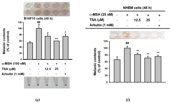
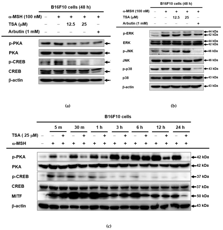
Theasinensin A (TSA) is a major group of catechin dimers mainly found in oolong tea and black tea. This compound is also manufactured with epigallocatechin gallate (EGCG) as a substrate and is refined after the enzyme reaction. In previous studies, TSA has been reported to be effective against inflammation. However, the effect of these substances on skin melanin formation remains unknown. In this study, we unraveled the role of TSA in melanogenesis using mouse melanoma B16F10 cells and normal human epidermal melanocytes (NHEMs) through reverse transcription polymerase chain reaction (RT-PCR), Western blotting analysis, luciferase reporter assay, and enzyme-linked immunosorbent assay analysis. TSA inhibited melanin formation and secretion in α-melanocyte stimulating hormone (α-MSH)-induced B16F10 cells and NHEMs. TSA down-regulated the mRNA expression of tyrosinase (Tyr), tyrosinase-related protein 1 (Tyrp1), and Tyrp2, which are all related to melanin formation in these cells. TSA was able to suppress the activities of certain proteins in the melanocortin 1 receptor (MC1R) signaling pathway associated with melanin synthesis in B16F10 cells: cyclic adenosine monophosphate (cAMP) response element-binding protein (CREB), protein kinase A (PKA), tyrosinase, and microphthalmia-associated transcription factor (MITF). We also confirmed α-MSH-mediated CREB activities through a luciferase reporter assay, and that the quantities of cAMP were reduced by TSA in the enzyme linked immunosorbent assay (ELISA) results. Based on these findings, TSA should be considered an effective inhibitor of hyperpigmentation.

Keywords: CREB; MC1R; cAMP; melanogenesis; theasinensin A.


Conflict of interest statement

The authors declare no conflict of interest.

Figures

Figure 1
Figure 1
Structure of theasinensin A (TSA). TSA was synthesized by the enzymatic reaction of epigallocatechin gallate (EGCG).
Figure 2
Figure 2
The antimelanogenesis effect of TSA in murine melanoma cells (B16F10). (a,b) B16F10 cells and NHEMs were treated with TSA (6.25–200 µM) for 48 h, and cell viability was analyzed using MTT assay (n = 5 from 3 independent experiments). (c) Mushroom tyrosinase was reacted with TSA (6.25–25 µM) or kojic acid (300 μM), and L-DOPA was added. Tyrosinase activity was analyzed by measuring absorbance at 475 nm (n = 4 from 3 replicates). (df) B16F10 cells and MHEMs were treated with TSA (12.5–25 µM) or arbutin (1 mM) for 48 h. Cell-cultured media were collected for melanin secretions by measuring absorbance at 470 nm (n = 4 from 3 independent experiments). The intracellular melanin contents were determined by measuring optical density at 450 nm with 3 replications. +: indicates treatment, −: indicates non-treatment. For all applicable experiments, statistical significance was evaluated using the Mann–Whitney U test. ## p < 0.01 compared with the normal group, * p < 0.05 compared with the control group, ** p < 0.01 compared with the control group.
Figure 2
Figure 2
The antimelanogenesis effect of TSA in murine melanoma cells (B16F10). (a,b) B16F10 cells and NHEMs were treated with TSA (6.25–200 µM) for 48 h, and cell viability was analyzed using MTT assay (n = 5 from 3 independent experiments). (c) Mushroom tyrosinase was reacted with TSA (6.25–25 µM) or kojic acid (300 μM), and L-DOPA was added. Tyrosinase activity was analyzed by measuring absorbance at 475 nm (n = 4 from 3 replicates). (df) B16F10 cells and MHEMs were treated with TSA (12.5–25 µM) or arbutin (1 mM) for 48 h. Cell-cultured media were collected for melanin secretions by measuring absorbance at 470 nm (n = 4 from 3 independent experiments). The intracellular melanin contents were determined by measuring optical density at 450 nm with 3 replications. +: indicates treatment, −: indicates non-treatment. For all applicable experiments, statistical significance was evaluated using the Mann–Whitney U test. ## p < 0.01 compared with the normal group, * p < 0.05 compared with the control group, ** p < 0.01 compared with the control group.
Figure 3
Figure 3
The effect on transcriptional events of melanogenesis. (a–d) The mRNA expression of Tyr, Tyrp1, and Tyrp2 in B16F10 or NHEMs treated with TSA (12.5–25 µM) or arbutin (1 mM) in response to α-MSH for 24 or 48 h were determined using RT-PCR and real-time PCR analyses. (e) Protein levels of tyrosinase and MITF in TSA (12.5–25 µM)- or arbutin (1 mM)-treated B16F10 cells, in response to α-MSH for 48 h of TSA, were determined using Western blotting analysis. For all applicable experiments, statistical significance was evaluated using the Mann–Whitney U test. All experiments were performed at least three times. +: indicates treatment, −: indicates non-treatment. ## p < 0.01 compared with the normal group, * p < 0.05 and ** p < 0.01 compared with the control group.
Figure 4
Figure 4
Regulation of the cAMP/CREB signaling pathway of TSA in B16F10 cells. (ac) Protein levels of various cyclic adenosine monophosphate (cAMP) signaling proteins and MAPK signaling proteins in B16F10 cells pretreated with TSA (12.5–25 µM) or arbutin (1 mM) in response to α-MSH at indicated time points were determined using Western blotting analysis. (d) Activation of CREB in B16F10 cells treated with TSA (12.5–25 µM) or arbutin (1 mM) was determined using the CREB-mediated luciferase system (n = 4 with three experimental replicates). (e) Levels of cAMP in B16F10 cells treated with TSA (12.5–25 µM) or arbutin (1 mM) were measured using an ELISA assay (n = 4). For all applicable experiments, statistical significance was evaluated using the Mann–Whitney U test. ## p < 0.01 compared with the normal group, * p < 0.05 and ** p < 0.01 compared with the control group.
Figure 4
Figure 4
Regulation of the cAMP/CREB signaling pathway of TSA in B16F10 cells. (ac) Protein levels of various cyclic adenosine monophosphate (cAMP) signaling proteins and MAPK signaling proteins in B16F10 cells pretreated with TSA (12.5–25 µM) or arbutin (1 mM) in response to α-MSH at indicated time points were determined using Western blotting analysis. (d) Activation of CREB in B16F10 cells treated with TSA (12.5–25 µM) or arbutin (1 mM) was determined using the CREB-mediated luciferase system (n = 4 with three experimental replicates). (e) Levels of cAMP in B16F10 cells treated with TSA (12.5–25 µM) or arbutin (1 mM) were measured using an ELISA assay (n = 4). For all applicable experiments, statistical significance was evaluated using the Mann–Whitney U test. ## p < 0.01 compared with the normal group, * p < 0.05 and ** p < 0.01 compared with the control group.
Figure 5
Figure 5
Mechanism of melanogenesis regulated by TSA. Treatment with TSA suppressed melanin Figure 1. and Tyrp2 through the downregulation of the cAMP pathway in α-MSH-treated B16F10 cells and NHEMs. α-MSH: α-melanocyte stimulating hormone, MC1R: melanocortin 1 receptor, AC: adenylyl cyclase, cAMP: cyclic adenosine monophosphate, PKA: protein kinase A, CREB: cAMP response element-binding protein, MAPK: mitogen-activated protein kinase, JNK: c-Jun-N-terminal kinase, ERK: extracellular signal-regulated kinase, MITF: microphthalmia-associated transcription factor, TRRP1: tyrosinase-related protein 1, and TYRP2: tyrosinase-related protein 1. Arrows indicate positive signal. Lines with theasinensin A indicate inhibitory signal.

References

    1. Qomaladewi N.P., Kim M.Y., Cho J.Y. Rottlerin Reduces cAMP/CREB-Mediated Melanogenesis via Regulation of Autophagy. Int. J. Mol. Sci. 2019;20:2081. doi: 10.3390/ijms20092081. - DOI - PMC - PubMed
    1. Zhu L., Lu Y., Yu W.G., Zhao X., Lu Y.H. Anti-photoageing and anti-melanogenesis activities of chrysin. Pharm. Biol. 2016;54:2692–2700. doi: 10.1080/13880209.2016.1179334. - DOI - PubMed
    1. Dabrowska A.K., Spano F., Derler S., Adlhart C., Spencer N.D., Rossi R.M. The relationship between skin function, barrier properties, and body-dependent factors. Skin Res. Technol. 2018;24:165–174. doi: 10.1111/srt.12424. - DOI - PubMed
    1. Gravitz L. Skin. Nature. 2018;563:S83. doi: 10.1038/d41586-018-07428-4. - DOI - PubMed
    1. Lee J.O., Kim E., Kim J.H., Hong Y.H., Kim H.G., Jeong D., Kim J., Kim S.H., Park C., Seo D.B., et al. Antimelanogenesis and skin-protective activities of Panax ginseng calyx ethanol extract. J. Ginseng Res. 2018;42:389–399. doi: 10.1016/j.jgr.2018.02.007. - DOI - PMC - PubMed