. 2021 Jul 14;22(14):7549.
doi: 10.3390/ijms22147549.

N-Acetylcysteine Added to Local Anesthesia Reduces Scar Area and Width in Early Wound Healing-An Animal Model Study

Affiliations

N-Acetylcysteine Added to Local Anesthesia Reduces Scar Area and Width in Early Wound Healing-An Animal Model Study

Wiktor Paskal et al. Int J Mol Sci. .

Abstract

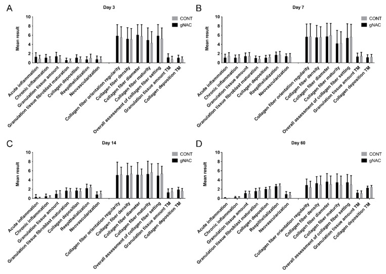
The aim of the study was to evaluate if a pre-incisional N-acetylcysteine (NAC) treatment altered the process of wound healing in a rat model. The dorsal skin of 24 Sprague-Dawley rats was incised in six locations. Before the incisions were made, skin was injected either with lidocaine and epinephrine (one side) or with these agents supplemented with 0.015%, 0.03%, or 0.045% NAC (contralaterally). Photographic documentation of the wound healing process was made at 11 time points. Rats were sacrificed 3, 7, 14, or 60 days after incision to excise scars for histological analysis. They included: Abramov scale scoring, histomorphometry analysis, and collagen fiber arrangement assessment. Skin pretreated with 0.03% NAC produced the shortest scars at all analyzed time points, though this result was statistically insignificant. At this NAC concentration the scars had smaller areas on the third day and were narrower on the day 4 compared with all the other groups (p < 0.05). On day 7, at the same concentration of NAC, the scars had a higher superficial concentration index (p = 0.03) and larger dermal proliferation area (p = 0.04). NAC addition to pre-incisional anesthetic solution decreased wound size and width at an early stage of scar formation at all concentrations; however, with optimal results at 0.03% concentration.

Keywords: N-Acetylcysteine; incision; local anesthesia additive; pretreatment; rat; skin; surgical; wound healing.


Conflict of interest statement

The authors declare no conflict of interest. The funders had no role in the design of the study; in the collection, analyses, or interpretation of data; in the writing of the manuscript, or in the decision to publish the results.

Figures

Figure 1
Figure 1
Video presents wound healing process captured with 11 time points of 60-days observation of a representative rat.
Figure 2
Figure 2
Graph represents results of planimetric measurements of photographed scars. (A) scar areas at days 1–60, (B) scar lengths at days 1–7, (C) scar mean width at days 1–60, (D) scar mean width at days 1–7, (E) scar lengths at day 1–60, (F) scar lengths at day 1–7. Mean results ± SD. * p < 0.05 (NAC30 vs. CONT).
Figure 3
Figure 3
Graph presents results of histological analysis with Abramov scale. Results of groups NAC15, NAC30, and NAC45 collectively analyzed as gNAC. (A) Day 3, (B) day 7, (C) day 14, (D) day 60. Mean values are shown. Error bars are SD. TM—Masson’s Trichrome stained images.
Figure 4
Figure 4
HE sections of representative samples from each group (NAC15, NAC30, NAC45, and CONT) at each harvest point. 1mm scale bar provided under each sample.
Figure 5
Figure 5
Graph summarizing differences in dermal proliferation area (A) and superficial contraction index (B) between NAC30 and CONT at day 7. Data expressed as mean ± SD, * p < 0.05.
Figure 6
Figure 6
Graph represents directional variance distribution of collagen fibers in proximal scar areas (A) and scars (B) on the 7th and 14th days, respectively, in gNAC vs. CONT groups. Values expressed as mean ± SD, * p < 0.05.
Figure 7
Figure 7
Representative results of automated collagen fiber arrangement analysis by Quinn et al. Heatmaps of collagen fiber density and average directional variance are presented along with Trichrome Masson’s staining of each group (NAC15, NAC30, NAC45, CONT) in four consecutive harvest points; 3rd, 7th, 14th, and 60th day. Quantitative analysis showed significant differences in the directional variance of collagen fibers surrounding the scar between the NAC groups versus CONT on the 7th and 14th day. 5mm scalebar provided under each time point.
Figure 8
Figure 8
Diagram of incision lines and injection points on the rat’s dorsum. x—pre-incisional injection points of intradermal administration (0.1 mL). Straight line—marked incision line, 1.5 cm long. Two 4-0 mattress sutures were placed between the injection points of each wound.
Figure 9
Figure 9
(A) A 3rd day wound with a surgical microscale (1 sqr–1 mm), (B) wound with marked area (green zone), length (blue line), and width (red lines) in ImageJ by a blinded researcher.
Figure 10
Figure 10
Diagram illustrates parameters measured in each HE section according to a mathematical model of wound healing proposed by Lemo et al. [15]. D—depth of the wound, DPA—dermal proliferation area, E—thickness of the epidermis, L—length of the re-epithelialization zone, N—thickness of the natural dermis, NE—thickness of the newly formed epidermis, S—distance between the borders of the wound, T—thickness of the connective tissue in the wound.
Figure 11
Figure 11
Diagram represents regions selected for automated collagen fiber analysis in MT-stained samples. Five areas were chosen, scar and four boxes 500 × 500 pixels of proximal and distant to scar area. S—scar zone, LD—left distant area, LP—left proximal area, RD—right distant area, RP—right proximal area.

References

    1. Rodrigues M., Kosaric N., Bonham C.A., Gurtner G.C. Wound Healing: A Cellular Perspective. Physiol. Rev. 2019;99:665–706. doi: 10.1152/physrev.00067.2017. - DOI - PMC - PubMed
    1. Landen N.X., Li D., Stahle M. Transition from inflammation to proliferation: A critical step during wound healing. Cell. Mol. Life Sci. 2016;73:3861–3885. doi: 10.1007/s00018-016-2268-0. - DOI - PMC - PubMed
    1. Becker D.E., Reed K.L. Local anesthetics: Review of pharmacological considerations. Anesth. Prog. 2012;59:90–101. doi: 10.2344/0003-3006-59.2.90. quiz 102–103. - DOI - PMC - PubMed
    1. Geers N.C., Zegel M., Huybregts J.G.J., Niessen F.B. The Influence of Preoperative Interventions on Postoperative Surgical Wound Healing in Patients Without Risk Factors: A Systematic Review. Aesthetic Surg. J. 2018;38:1237–1249. doi: 10.1093/asj/sjy074. - DOI - PubMed
    1. Zampieri N., Zuin V., Burro R., Ottolenghi A., Camoglio F.S. A prospective study in children: Pre- and post-surgery use of vitamin E in surgical incisions. J. Plast. Reconstr. Aesthetic Surg. 2010;63:1474–1478. doi: 10.1016/j.bjps.2009.08.018. - DOI - PubMed