Review
. 2021 Jul 16;22(14):7618.
doi: 10.3390/ijms22147618.

Innate Lymphoid Cells in Intestinal Homeostasis and Inflammatory Bowel Disease

Affiliations
Review

Innate Lymphoid Cells in Intestinal Homeostasis and Inflammatory Bowel Disease

Angela Saez et al. Int J Mol Sci. .

Abstract

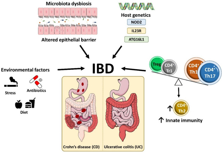
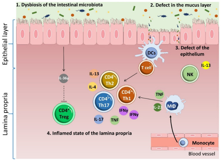
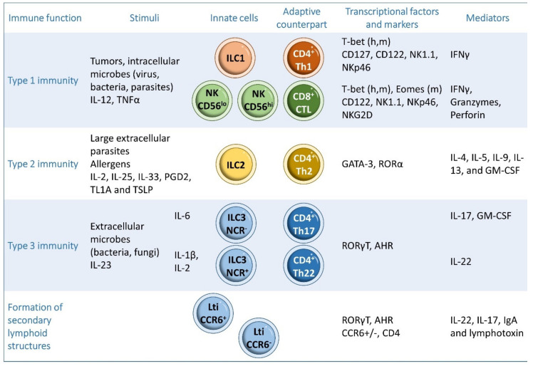
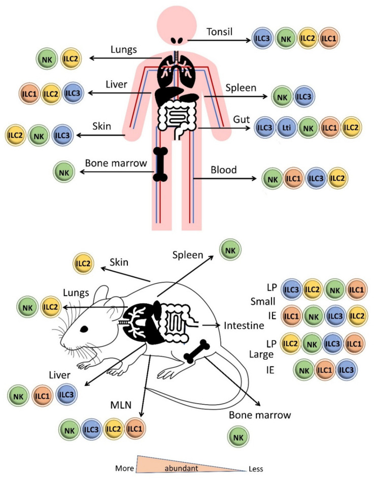
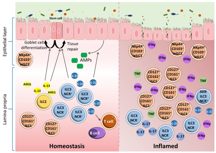
Inflammatory bowel disease (IBD) is a heterogeneous state of chronic intestinal inflammation of unknown cause encompassing Crohn's disease (CD) and ulcerative colitis (UC). IBD has been linked to genetic and environmental factors, microbiota dysbiosis, exacerbated innate and adaptive immunity and epithelial intestinal barrier dysfunction. IBD is classically associated with gut accumulation of proinflammatory Th1 and Th17 cells accompanied by insufficient Treg numbers and Tr1 immune suppression. Inflammatory T cells guide innate cells to perpetuate a constant hypersensitivity to microbial antigens, tissue injury and chronic intestinal inflammation. Recent studies of intestinal mucosal homeostasis and IBD suggest involvement of innate lymphoid cells (ILCs). These lymphoid-origin cells are innate counterparts of T cells but lack the antigen receptors expressed on B and T cells. ILCs play important roles in the first line of antimicrobial defense and contribute to organ development, tissue protection and regeneration, and mucosal homeostasis by maintaining the balance between antipathogen immunity and commensal tolerance. Intestinal homeostasis requires strict regulation of the quantity and activity of local ILC subpopulations. Recent studies demonstrated that changes to ILCs during IBD contribute to disease development. A better understanding of ILC behavior in gastrointestinal homeostasis and inflammation will provide valuable insights into new approaches to IBD treatment. This review summarizes recent research into ILCs in intestinal homeostasis and the latest advances in the understanding of the role of ILCs in IBD, with particular emphasis on the interaction between microbiota and ILC populations and functions.

Keywords: inflammatory bowel disease; innate lymphoid cells; intestinal homeostasis.


Conflict of interest statement

The authors declare no conflict of interest.

Figures

Figure 1
Figure 1
Etiology of inflammatory bowel disease. IBD is a heterogeneous state of chronic intestinal inflammation comprising two main clinical phenotypes, Crohn’s disease (CD) and ulcerative colitis (UC), which are distinguished by their symptoms, disease location and histopathological features. IBD arises from the interplay between environment factors, the gut microbiota and immunological factors in genetically susceptible individuals, which promotes intestinal barrier dysfunction, tissue damage and dysregulated innate and adaptive immune responses.
Figure 2
Figure 2
Pathophysiology of inflammatory bowel disease. Multiple factors contribute to IBD. Changes in the microbiota accompanied by thinning of the mucus layer induces a barrier breach that results in defects in the epithelium. Crossing of the barrier by microbiota components induces DC and macrophage activation, which induces infiltration of the intestinal tissue by inflammatory CD4 T cells. IBD patients have an increased content of proinflammatory Th1 and Th17 cells. This infiltration is accompanied by an increase in Th2 cell numbers and insufficient numbers of immune suppressing cells, such as Tregs. Inflammatory T cells guide the function of cells with an innate immune role, such as epithelial cells, fibroblasts and phagocytes, thus stimulating a constant hyperresponsiveness to microbial antigens and causing tissue injury and chronic intestinal inflammation.
Figure 3
Figure 3
Classification of ILCs. ILCs can be classified into three subgroups: type 1 ILCs, including natural killer (NK) cells and ILC1s, type 2 ILCs (ILC2s) and type 3 ILCs (ILC3s). h: human, m: mouse.
Figure 4
Figure 4
Distribution of ILCs in the body in human and mouse. NKs are circulating cells that are mainly found in the systemic circulation, cord blood, bone marrow, spleen, lungs and throughout the human gut. ILC1s are tissue-resident cells that mainly reside in intestinal tissues and tonsils but are also located in the liver, salivary glands, uterus and thymus. ILC2s are mainly found in adipose tissue, mesenteric lymph nodes, lungs, skin and tonsils. ILC3s are mainly present in mucosal tissue and at low levels in the spleen and liver. LTis cells are predominantly located in intestinal and lymphoid tissues, whereas NCR+ ILC3s and NCR ILC3s are more prominent in the skin and intestinal lamina propria. (LP: Lamina propria; IE: intraepithelial).
Figure 5
Figure 5
ILCs, homeostasis, and inflammatory bowel disease. There are two major subpopulations of ILC1s in the human gut: lamina propria ILC1s (CD161+ CD127+) and intraepithelial ILC1s (NKp44+ CD103+ CD127). ILC2s are present in lower numbers than ILC1s and ILC3s. ILC2s contribute to epithelial barrier maintenance through the production of IL-13, which promotes the differentiation of intestinal epithelial stem cells toward turf cells and goblet cells. ILC2s also maintain intestinal epithelia homeostasis through the production of amphiregulin (AREG). ILC3s maintain gastrointestinal tract homeostasis by producing IL-22. This cytokine activates intestinal epithelial cells to produce antimicrobial peptides, enhances the renewal of epithelial cells, promotes tissue repair and modulates the homeostasis of adaptive immunity. Alteration of the ILC subset profile disturbs intestinal homeostasis and leads to inflammation in the gut. ILC3s and ILC1s are involved in the induction of inflammation and are closely related to IBD pathogenesis. This conclusion is supported by the amplification of intraepithelial IFN-γ-producing ILC1s in response to IL-12 and IL-15 in the gut of CD patients. This increase is accompanied by a reduction in NCR+ ILC3s, enhancing disease severity.
Figure 6
Figure 6
Microbiota-ILC3 interactions in intestinal homeostasis. Some SCFAs are recognized by the receptor FFAR2 on ILC3s, triggering IL-22 cytokine production by stimulating the AKT–STAT3 and ERK–STAT3 signaling pathways. TLR2 stimulation in human ILC3s promotes the production of IL-22, IL-13 and IL-5 upon activation of signaling via NF-κB and JAK (not shown). Stimulation of the TLR5receptor on dendritic cells (DCs) with the bacterial protein flagellin promotes the production of IL-23 and IL-1b, which induces production of IL-22, and IL-2 and GM-CSF by ILC3s, respectively. In human ILC3s, ligation of the natural cytotoxicity receptor NKp44 promotes the production of TNFα by ILC3s and, in combination with IL-1, IL-17 and IL-23, enhances ILC3 production of IL-22, GM-CSF, IL-2 and TNFα in a mechanism mediated by NF-κB and Nuclear factor of activated T-cells (NFAT) signaling. NKp44 receptor activation also increases the proportion of IL-22-producing ILC3s. IL-22 facilitates intestinal homeostasis by preservation of epithelial barrier function and promoting the secretion of antibacterial peptides (AMPs). IL-2 and GM-CSF contributes to intestinal tolerance mediated by Tregs. Tregs prevent ILC3-associated colitis by inhibiting IL-23- and IL-1β-induced IL-22 production.
Figure 7
Figure 7
Influence of microbiota in other ILCs in intestinal homeostasis. (a) SCFAs produced by the commensal gut microbiota support optimal proliferation of ILC1, ILC2 and ILC3 populations by regulating G protein-coupled receptors (GPCRs). (b) In the absence of commensal bacteria, NK cells have reduced cytotoxicity and IFN-γ production. Colonization of germ-free mice with commensals increases NK cytotoxicity through the effect of dendritic cells and macrophage-derived type-I interferons on IL-15, which promotes NK cell terminal maturation. (c) LTi cells produce cryptopatches, which are transformed into isolated lymphoid follicles in a microbiota-dependent manner, supporting the production of intestinal IgA. (d) The gut microbiota enhances the expression of T-bet in ILCs. (e) The proportion of ILC2s in the gut is increased in the absence of commensal microbiota. (f) The microbiota regulates ILC2 function in the gut by promoting the release of IL-25, which drives ILC2s to improve intestinal barrier function.
Figure 8
Figure 8
ILC plasticity. Prolonged exposure of ILC3s to the type 1 polarizing cytokines IL-2, IL-12 and IL-15 induces their conversion to CD127+ ILC1 cells, with downregulation of RORC, upregulation of TBX21 and IFN-γ production. Reverse differentiation of ILC1s to ILC3s is driven by IL-23, IL-2 and IL-1β. Moreover, CD14 DCs mediate the transformation of ILC1s into ILC3s in vivo by promoting synthesis of c-kit and NKp44 in ILC1s, whereas CD14+ DCs are implicated in the transformation of NCR+ ILC3s into ILC1s. IL-23 and IL-1β stimulation induces the conversion of NCR ILC3s into the more proinflammatory NCR+ ILC3 subset through T-bet upregulation. The reverse differentiation from NCR+ ILC3s to NCR ILC3s is promoted by high levels of TGF-β. Removal of AHR enriches the effect of intestinal ILC2s, whereas increased AHR expression restrains ILC2 function while increasing ILC3 function. Treatment with IL-12 and IL-18 reduces IL-13 expression in ILC2s, shifting them to an IL-13 IFN-γ+ ILC1 phenotype. ILC2-derived ILC1s can revert to ILC2s in response to IL-4. NK cells (CD49a CD49b+ Eomes+) differentiate into intermediate type 1 innate lymphoid (intILC1, CD49a+ CD49b+ Eomes+) populations and ILC1s (CD49a+ CD49b Eomesint) in response to cytokine and TGF-β signaling.

References

    1. Satsangi J., Silverberg M.S., Vermeire S., Colombel J.F. The Montreal classification of inflammatory bowel disease: Controversies, consensus, and implications. Gut. 2006;55:749–753. doi: 10.1136/gut.2005.082909. - DOI - PMC - PubMed
    1. Global burden of inflammatory bowel disease 2017 Collaborators The global, regional, and national burden of inflammatory bowel disease in 195 countries and territories, 1990–2017: A systematic analysis for the Global Burden of Disease Study 2017. Lancet Gastroenterol. Hepatol. 2020;5:17–30. doi: 10.1016/S2468-1253(19)30333-4. - DOI - PMC - PubMed
    1. Peloquin J.M., Goel G., Villablanca E.J., Xavier R.J. Mechanisms of pediatric inflammatory bowel disease. Annu. Rev. Immunol. 2016;34:31–64. doi: 10.1146/annurev-immunol-032414-112151. - DOI - PubMed
    1. Liu T.C., Stappenbeck T.S. Genetics and pathogenesis of inflammatory bowel disease. Annu. Rev. Pathol. 2016;11:127–148. doi: 10.1146/annurev-pathol-012615-044152. - DOI - PMC - PubMed
    1. Torres J., Mehandru S., Colombel J.F., Peyrin-Biroulet L. Crohn’s disease. Lancet. 2017;389:1741–1755. doi: 10.1016/S0140-6736(16)31711-1. - DOI - PubMed

MeSH terms

LinkOut - more resources