Review
. 2021 Jul 17;22(14):7651.
doi: 10.3390/ijms22147651.

Magnetic Particle Imaging: Current and Future Applications, Magnetic Nanoparticle Synthesis Methods and Safety Measures

Affiliations
Review

Magnetic Particle Imaging: Current and Future Applications, Magnetic Nanoparticle Synthesis Methods and Safety Measures

Caroline Billings et al. Int J Mol Sci. .

Abstract

Magnetic nanoparticles (MNPs) have a wide range of applications; an area of particular interest is magnetic particle imaging (MPI). MPI is an imaging modality that utilizes superparamagnetic iron oxide particles (SPIONs) as tracer particles to produce highly sensitive and specific images in a broad range of applications, including cardiovascular, neuroimaging, tumor imaging, magnetic hyperthermia and cellular tracking. While there are hurdles to overcome, including accessibility of products, and an understanding of safety and toxicity profiles, MPI has the potential to revolutionize research and clinical biomedical imaging. This review will explore a brief history of MPI, MNP synthesis methods, current and future applications, and safety concerns associated with this newly emerging imaging modality.

Keywords: magnetic nanoparticles; magnetic particle imaging; nanoparticle safety; superparamagnetic iron oxide.


Conflict of interest statement

The authors declare no conflict of interest.

Figures

Figure 1
Figure 1
MRI/MPI image of nanoparticle distribution from a mouse model (a) which shows improvement of MPI signal intensity using CREKA peptide functionalization of iron oxide nanoparticles (b). *** p < 0.001, IOs-CREKA NPs or IOs compared with Vivotrax group; # p < 0.05, IOs versus IO-CREKA NPs.Reprinted with permission from Du et al. [54].
Figure 2
Figure 2
Tracers of SPIO bolus passing through the stroked brain of a mouse. Slices of fused 3D MPI data at several times are shown for the coronal, sagittal, and transverse planes. (Red arrows: occluded and non-occluded common/internal carotid. Blue arrow: the basilar artery). MR dynamic susceptibility contrast (DSC) images are shown for comparison (bottom row). Reprinted with permission from Ludewig et al. [72].
Figure 3
Figure 3
Generation and clearance of the extracellular SPIONs in the myocardium. Reprinted from Huang et al. [90] following the Creative Commons Attribution (CC BY) license (http://creativecommons.org/licenses/by/4.0/) (accessed on 4 March 2021).
Figure 4
Figure 4
Schematic representation of MRI gene reporter integration for in vivo detection of cell fate decisions. CEST (chemical exchange of saturated magnetization), TfR (transferrin receptor). Reprinted from Vandsburger [92] following the Creative Commons Attribution (CC BY) license (http://creativecommons.org/licenses/by/4.0/) (accessed on 6 March 2021).
Figure 5
Figure 5
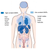
Representation of biodistribution of MNPs in organs and tissues. Unedited image obtained from Zamay et al. [104] following the Creative Commons Attribution (CC BY) license (http://creativecommons.org/licenses/by/4.0/). (accessed on 20 April 2021).
Figure 6
Figure 6
Cytotoxicity analysis of human fibroblast (HFF-2, a) and human embryonic kidney (HEK-293, b) cell lines with varying concentrations of SPIONs. Unedited image obtained from Nosrati et al. [110] following the Creative Commons License, (http://creativecommons.org/licenses/by/4.0/). (accessed on 20 April 2021).

References

    1. Bulte J.W.M. Superparamagnetic iron oxides as MPI tracers: A primer and review of early applications. Adv. Drug. Deliv. Rev. 2019;138:293–301. doi: 10.1016/j.addr.2018.12.007. - DOI - PMC - PubMed
    1. Sun C., Lee J.S., Zhang M. Magnetic nanoparticles in MR imaging and drug delivery. Adv. Drug. Deliv. Rev. 2008;60:1252–1265. doi: 10.1016/j.addr.2008.03.018. - DOI - PMC - PubMed
    1. Lu C., Han L., Wang J., Wan J., Song G., Rao J. Engineering of magnetic nanoparticles as magnetic particle imaging tracers. Chem. Soc. Rev. 2021 doi: 10.1039/D0CS00260G. - DOI - PubMed
    1. Knopp T., Gdaniec N., Moddel M. Magnetic particle imaging: From proof of principle to preclinical applications. Phys. Med. Biol. 2017;62:R124–R178. doi: 10.1088/1361-6560/aa6c99. - DOI - PubMed
    1. Williams H. The application of magnetic nanoparticles in the treatment and monitoring of cancer and infectious diseases. Biosci. Horiz. Int. J. Stud. Res. 2017;10:1–10. doi: 10.1093/biohorizons/hzx009. - DOI

MeSH terms