. 2021 Jul 13;26(14):4247.
doi: 10.3390/molecules26144247.

High Surface Area Mesoporous Silica Nanoparticles with Tunable Size in the Sub-Micrometer Regime: Insights on the Size and Porosity Control Mechanisms

Affiliations

High Surface Area Mesoporous Silica Nanoparticles with Tunable Size in the Sub-Micrometer Regime: Insights on the Size and Porosity Control Mechanisms

Federica Rizzi et al. Molecules. .

Abstract

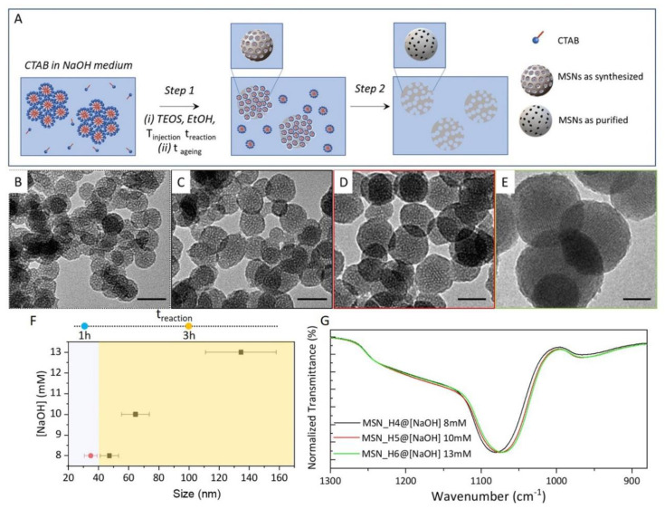
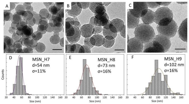
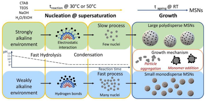
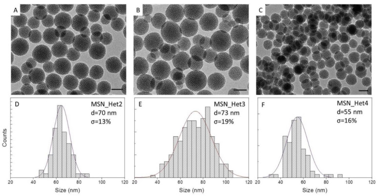
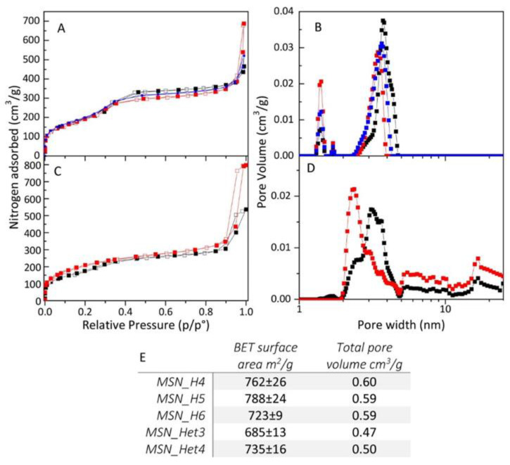
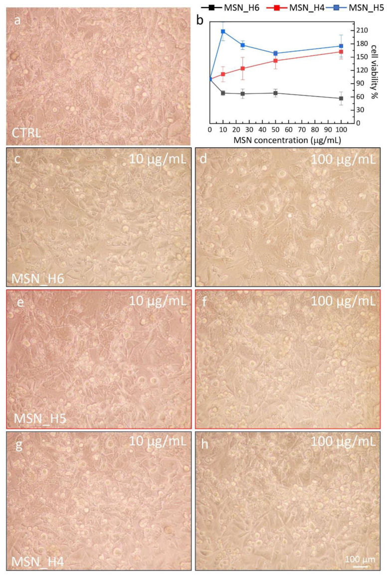
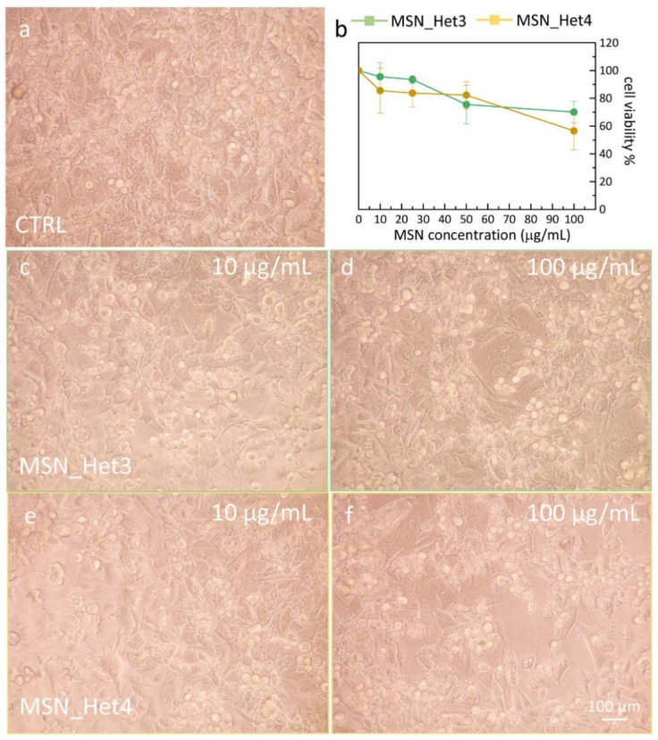
Mesoporous silica nanostructures (MSNs) attract high interest due to their unique and tunable physical chemical features, including high specific surface area and large pore volume, that hold a great potential in a variety of fields, i.e., adsorption, catalysis, and biomedicine. An essential feature for biomedical application of MSNs is limiting MSN size in the sub-micrometer regime to control uptake and cell viability. However, careful size tuning in such a regime remains still challenging. We aim to tackling this issue by developing two synthetic procedures for MSN size modulation, performed in homogenous aqueous/ethanol solution or two-phase aqueous/ethyl acetate system. Both approaches make use of tetraethyl orthosilicate as precursor, in the presence of cetyltrimethylammonium bromide, as structure-directing agent, and NaOH, as base-catalyst. NaOH catalyzed syntheses usually require high temperature (>80 °C) and large reaction medium volume to trigger MSN formation and limit aggregation. Here, a successful modulation of MSNs size from 40 up to 150 nm is demonstrated to be achieved by purposely balancing synthesis conditions, being able, in addition, to keep reaction temperature not higher than 50 °C (30 °C and 50 °C, respectively) and reaction mixture volume low. Through a comprehensive and in-depth systematic morphological and structural investigation, the mechanism and kinetics that sustain the control of MSNs size in such low dimensional regime are defined, highlighting that modulation of size and pores of the structures are mainly mediated by base concentration, reaction time and temperature and ageing, for the homogenous phase approach, and by temperature for the two-phase synthesis. Finally, an in vitro study is performed on bEnd.3 cells to investigate on the cytotoxicity of the MNSs.

Keywords: colloidal synthesis; high specific surface area; mesoporous silica nanoparticles.


Conflict of interest statement

The authors declare no conflict of interest.

Figures

Figure 1
Figure 1
(A) Sketch of the synthetic MSNs in homogeneous solution. Step 1: (i) EtOH and TEOS are added to CTAB micellar aqueous solution at alkaline pH by NaOH at injection temperature, Tinjection, the reaction mixture is let stirring for different reaction time (treaction) then (ii) the temperature is decreased at room temperature (tageing) to allow MSNs growth. Step 2: surfactant template removal; (BE) TEM micrographs (scale bar 50 nm) of MSNs (see also Table 1) MSN_H3 (B), MSN_H4 (C), MSN_H5 (D) and MSN_H6 (E); (F) Scatter plot of MSN average size, including size distribution as function of NaOH concentration and treaction; (G) FTIR spectra recorded in ATR mode in the 1300–880 cm−1 range of MSNs samples (panel (F)).
Figure 2
Figure 2
TEM micrographs ((AC), scale bar 50 nm) and the corresponding size distribution statistical analysis (DF) of MSN_H7 (A,D), MSN_H8 (B,E) and MSN_H9 (D,F).
Figure 3
Figure 3
Scheme of the pH-dependent nucleation and growth of the MSNs, prepared in homogenous solution. pH value controls the kinetic of hydrolysis and condensation during the nucleation at high reaction temperature (treaction), and the growth step at room temperature.
Figure 4
Figure 4
TEM micrographs (AC, scale bar 50 nm) and the corresponding size distribution statistical analysis (DF) of MSNs prepared by injecting TEOS 4.47 mmol (1 mL) at Tinjection = 50 °C ((A,B,D,E) MSN_Het2 and MSN_Het3 samples) or Tinjecttion = 30 °C ((C,F) MSN_Het4 sample) to 50 mL of H2O/Ethyl acetate (50:2 v/v), [CTAB] = 5 mM and [NaOH] 5 mM ((A,D) MSN_Het2) and 13 mM ((B,C,E,F), MSN_Het3, MSN_Het 4). treaction 3 h and tageing 24 h.
Figure 5
Figure 5
(AC) N2 adsorption-desorption isotherms and (B,D) pore width distributions by NLDFT of MSN_H4 (panel A, B red line), MSN_H5 (panel A, B blue line), MSN_H6 (panel A, B black line), MSN_Het3 (panel C, D, black line), MSN_Het4 (panel C, D red line); (E) table of MSNs samples surface area and pore volume.
Figure 6
Figure 6
(a,ch) Representative phase contrast optical images showing the morphology of bEnd.3 (20× magnification) and of the control (CTRL, 100%) after 24 h treatment with the MSN samples as labeled, prepared by the homogenous solution approaches, at the indicated concentrations. (b) Graph of the cell viability, determined by the MTT test, expressed as percentage of surviving cells in comparison with control (CTRL, 100%) represented by untreated bEnd.3 cells in serum-free DMEM.
Figure 7
Figure 7
(a,cf) Representative phase contrast optical images showing the morphology of bEnd.3 (20× magnification) and of the control (CTRL, 100%) after 24 h treatment with MSN samples as labeled, prepared by the two-phase approaches, at the indicated concentrations. (b) Graph of the cell viability, determined by the MTT test, expressed as percentage of surviving cells in comparison with control (CTRL, 100%) represented by untreated bEnd.3 cells in serum-free DMEM.

References

    1. Jarmolińska S., Feliczak-Guzik A., Nowak I. Synthesis, Characterization and Use of Mesoporous Silicas of the Following Types SBA-1, SBA-2, HMM-1 and HMM-2. Materials. 2020;13:4385. doi: 10.3390/ma13194385. - DOI - PMC - PubMed
    1. Olivieri F., Castaldo R., Cocca M., Gentile G., Lavorgna M. Mesoporous silica nanoparticles as carriers of active agents for smart anticorrosive organic coatings: A critical review. Nanoscale. 2021;13:9091–9111. doi: 10.1039/D1NR01899J. - DOI - PubMed
    1. Pal N., Lee J.-H., Cho E.-B. Recent Trends in Morphology-Controlled Synthesis and Application of Mesoporous Silica Nanoparticles. Nanomaterials. 2020;10:2122. doi: 10.3390/nano10112122. - DOI - PMC - PubMed
    1. von Baeckmann C., Guillet-Nicolas R., Renfer D., Kählig H., Kleitz F. A Toolbox for the Synthesis of Multifunctionalized Mesoporous Silica Nanoparticles for Biomedical Applications. ACS Omega. 2018;3:17496–17510. doi: 10.1021/acsomega.8b02784. - DOI - PMC - PubMed
    1. Jafari S., Derakhshankhah H., Alaei L., Fattahi A., Varnamkhasti B.S., Saboury A.A. Mesoporous silica nanoparticles for therapeutic/diagnostic applications. Biomed. Pharmacother. 2019;109:1100–1111. doi: 10.1016/j.biopha.2018.10.167. - DOI - PubMed