

Recent Advancements in 3D Printing of Polysaccharide Hydrogels in Cartilage Tissue Engineering
- PMID: 34300896
- PMCID: PMC8305911
- DOI: 10.3390/ma14143977
Recent Advancements in 3D Printing of Polysaccharide Hydrogels in Cartilage Tissue Engineering
Abstract
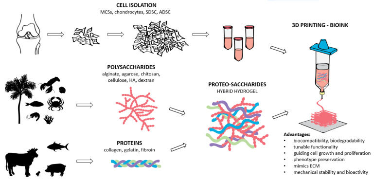
The application of hydrogels coupled with 3-dimensional (3D) printing technologies represents a modern concept in scaffold development in cartilage tissue engineering (CTE). Hydrogels based on natural biomaterials are extensively used for this purpose. This is mainly due to their excellent biocompatibility, inherent bioactivity, and special microstructure that supports tissue regeneration. The use of natural biomaterials, especially polysaccharides and proteins, represents an attractive strategy towards scaffold formation as they mimic the structure of extracellular matrix (ECM) and guide cell growth, proliferation, and phenotype preservation. Polysaccharide-based hydrogels, such as alginate, agarose, chitosan, cellulose, hyaluronan, and dextran, are distinctive scaffold materials with advantageous properties, low cytotoxicity, and tunable functionality. These superior properties can be further complemented with various proteins (e.g., collagen, gelatin, fibroin), forming novel base formulations termed "proteo-saccharides" to improve the scaffold's physiological signaling and mechanical strength. This review highlights the significance of 3D bioprinted scaffolds of natural-based hydrogels used in CTE. Further, the printability and bioink formation of the proteo-saccharides-based hydrogels have also been discussed, including the possible clinical translation of such materials.
Keywords: 3D (bio)printing; cartilage tissue engineering; hydrogels; polysaccharides; proteins; proteo-saccharides.
Conflict of interest statement
The authors declare no conflict of interest.
Figures
References
-
- Eftekhari A., Maleki Dizaj S., Sharifi S., Salatin S., Rahbar Saadat Y., Zununi Vahed S., Samiei M., Ardalan M., Rameshrad M., Ahmadian E., et al. The Use of Nanomaterials in Tissue Engineering for Cartilage Regeneration; Current Approaches and Future Perspectives. Int. J. Mol. Sci. 2020;21:536. doi: 10.3390/ijms21020536. - DOI - PMC - PubMed
-
- Del Bakhshayesh A.R., Asadi N., Alihemmati A., Tayefi Nasrabadi H., Montaseri A., Davaran S., Saghati S., Akbarzadeh A., Abedelahi A. An Overview of Advanced Biocompatible and Biomimetic Materials for Creation of Replacement Structures in the Musculoskeletal Systems: Focusing on Cartilage Tissue Engineering. J. Biol. Eng. 2019;13:85. doi: 10.1186/s13036-019-0209-9. - DOI - PMC - PubMed
Publication types
Grants and funding
LinkOut - more resources
Full Text Sources
