

Validity and reliability of the medial temporal lobe atrophy scale in a memory clinic population
- PMID: 34301202
- PMCID: PMC8305846
- DOI: 10.1186/s12883-021-02325-2
Validity and reliability of the medial temporal lobe atrophy scale in a memory clinic population
Abstract
Background: Visual rating of medial temporal lobe atrophy (MTA) is often performed in conjunction with dementia workup. Most prior studies involved patients with known or probable Alzheimer's disease (AD). This study investigated the validity and reliability of MTA in a memory clinic population.
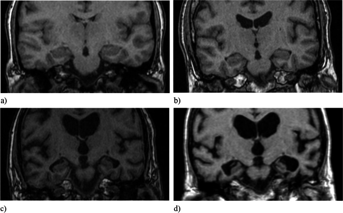
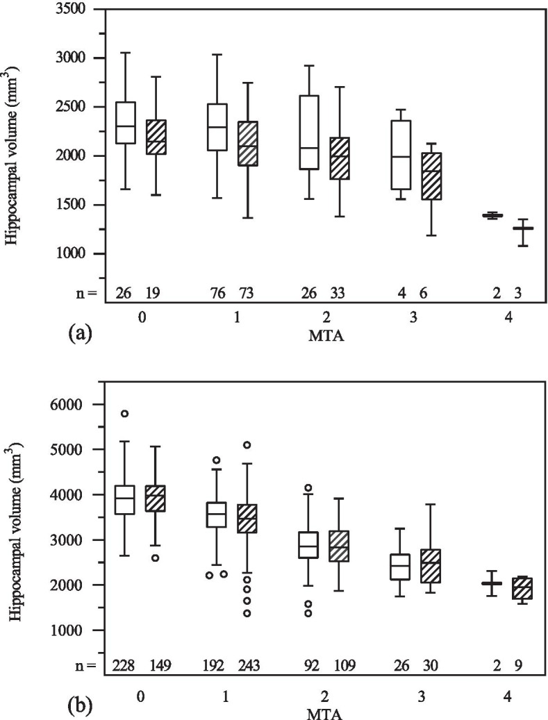
Methods: MTA was rated in 752 MRI examinations, of which 105 were performed in cognitively healthy participants (CH), 184 in participants with subjective cognitive impairment, 249 in subjects with mild cognitive impairment, and 214 in patients with dementia, including AD, subcortical vascular dementia and mixed dementia. Hippocampal volumes, measured manually or using FreeSurfer, were available in the majority of cases. Intra- and interrater reliability was tested using Cohen's weighted kappa. Correlation between MTA and quantitative hippocampal measurements was ascertained with Spearman's rank correlation coefficient. Moreover, diagnostic ability of MTA was assessed with receiver operating characteristic (ROC) analysis and suitable, age-dependent MTA thresholds were determined.
Results: Rater agreement was moderate to substantial. MTA correlation with quantitative volumetric methods ranged from -0.20 (p< 0.05) to -0.68 (p < 0.001) depending on the quantitative method used. Both MTA and FreeSurfer are able to distinguish dementia subgroups from CH. Suggested age-dependent MTA thresholds are 1 for the age group below 75 years and 1.5 for the age group 75 years and older.
Conclusions: MTA can be considered a valid marker of medial temporal lobe atrophy and may thus be valuable in the assessment of patients with cognitive impairment, even in a heterogeneous patient population.
Keywords: Alzheimer’s disease; Atrophy; Dementia; Magnetic resonance imaging; Medial temporal lobe atrophy (MTA); Mild cognitive impairment.
© 2021. The Author(s).
Conflict of interest statement
The authors declare that they have no competing interests.
Figures
References
MeSH terms
LinkOut - more resources
Full Text Sources
Medical
