. 2021 Aug 1;35(15-16):1109-1122.
doi: 10.1101/gad.347344.120. Epub 2021 Jul 22.

miR-200 deficiency promotes lung cancer metastasis by activating Notch signaling in cancer-associated fibroblasts

Affiliations

miR-200 deficiency promotes lung cancer metastasis by activating Notch signaling in cancer-associated fibroblasts

Bin Xue et al. Genes Dev. .

Abstract

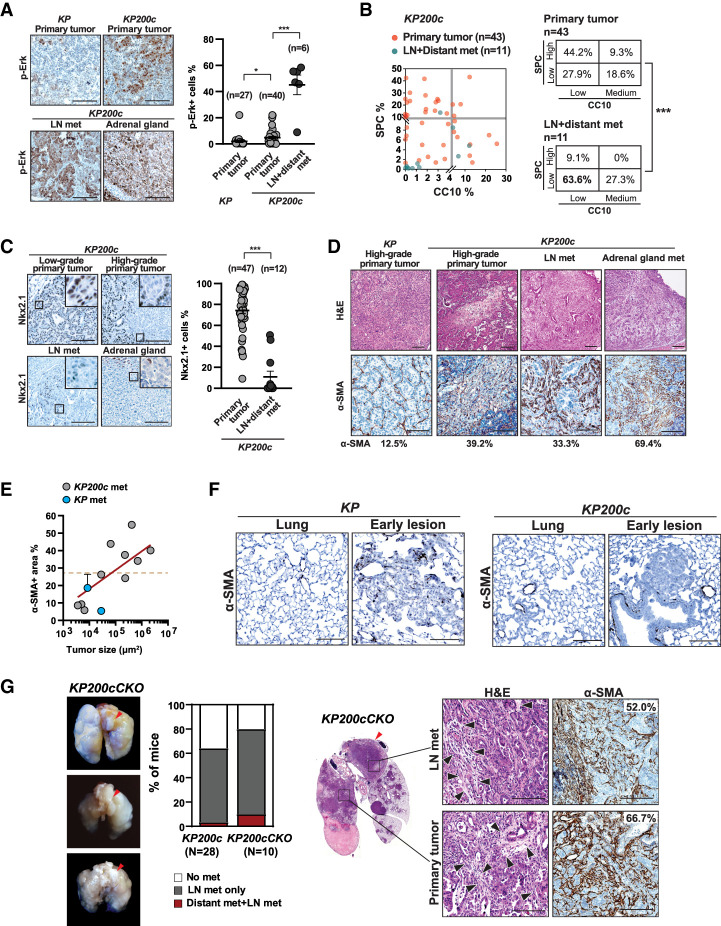
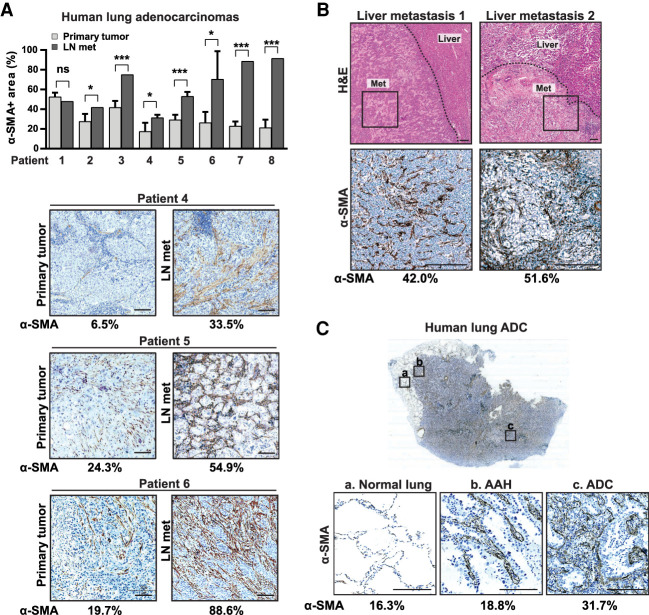
Lung adenocarcinoma, the most prevalent lung cancer subtype, is characterized by its high propensity to metastasize. Despite the importance of metastasis in lung cancer mortality, its underlying cellular and molecular mechanisms remain largely elusive. Here, we identified miR-200 miRNAs as potent suppressors for lung adenocarcinoma metastasis. miR-200 expression is specifically repressed in mouse metastatic lung adenocarcinomas, and miR-200 decrease strongly correlates with poor patient survival. Consistently, deletion of mir-200c/141 in the KrasLSL-G12D/+ ; Trp53flox/flox lung adenocarcinoma mouse model significantly promoted metastasis, generating a desmoplastic tumor stroma highly reminiscent of metastatic human lung cancer. miR-200 deficiency in lung cancer cells promotes the proliferation and activation of adjacent cancer-associated fibroblasts (CAFs), which in turn elevates the metastatic potential of cancer cells. miR-200 regulates the functional interaction between cancer cells and CAFs, at least in part, by targeting Notch ligand Jagged1 and Jagged2 in cancer cells and inducing Notch activation in adjacent CAFs. Hence, the interaction between cancer cells and CAFs constitutes an essential mechanism to promote metastatic potential.

Keywords: Jag1; Jag2; cancer-associated fibroblasts; lung cancer; metastasis; miR-141; miR-200; miR-200c; miRNA; microenvironment.


Figures

Figure 1.
Figure 1.
miR-200 deficiency induces rapid metastasis in KP lung cancer model. (A) Cartoon illustrating isolation of tdTomato+ cancer cells from primary and metastatic KPT lung adenocarcinomas for miRNA profiling. (B) miR-200 miRNAs are strongly down-regulated in distant metastases in the KPT mouse lung adenocarcinoma model. A heat map is shown for the most differentially expressed miRNAs between seven KPT primary lung tumors and four distant metastases (P < 0.05; expression level ≥−20). (Red text) miR-200 miRNAs. (C) The five miR-200 miRNAs reside in two genomic loci and segregate into two subfamilies based on seed sequences. Red box, seed sequences. (D) miR-200 miRNAs are strongly down-regulated in metastatic KPT tumors. Real-time PCR detected miR-200 expression in KP early stage primary tumors (N = 2) and late stage primary tumors (n = 10), but not in KP metastases (n = 7). Error bars indicate SEM. Late stage primary tumors versus metastases. miR-200a: (**) P = 0.0052, t = 3.268, df = 15; miR-429: (*) P = 0.0113, t = 2.887, df = 15; miR-141: (**) P = 0.01, t = 2.047, df = 15; miR-200c: (**) P = 0.0068, t = 3.395, df = 10. All statistical analyses were performed using unpaired, two tailed, Student's t-test. (E) Decreased total MIR-200 expression human lung adenocarcinomas is associated with poor patient survival in the TCGA data (LUAD). A Kaplan-Meier plot compares patient survival between two cohort of patients, with either high (N = 410) or low (N = 22) expression of all five MIR-200 miRNAs. (**) P = 0.00188, log rank test. (F) miR-200c/141 deficiency in the KP200c model significantly reduces overall survival. A Kaplan-Meier plot compares survival of KP and KP200c mice after Adeno-Cre administration (5 × 10⁶ Ad-Cre PFU/mouse), demonstrating a significant acceleration of tumor progression in KP200c mice. (***) P = 0.0004, log rank test. (G) miR-200c/141 deficiency in KP200c mice induces a significant increase in tumor progression. Representative H&E staining of lung sections are shown for three pairs of KP and KP200c mice collected at 90 d after tumor initiation. (Red arrows) Lymph node metastases. (H) KP200c primary tumors are characterized by desmoplastic stroma (blue arrows) and pleomorphic nuclei in cancer cells (red arrows). Representative images are shown for a pair of late stage KP and KP200c primary tumors collected at terminal stage. Scale bars, 100 μm. (I) miR-200 deficiency leads to a significant increase in tumor burden, tumor size, and tumor grade in KP200c mice. (Left) Tumor burden is quantified as the percentage of tumor area versus total lung area. Error bars indicate SEM. (***) P < 0.001. (N) Animal number, unpaired two-tailed Student's t-test, t = 8.199, df = 21. (Middle) Tumor size was measured by tumor area. Error bars indicate SEM. (*) P = 0.0245. (n) Tumor number, unpaired two-tailed Student's t-test, t = 2.278, df = 118. (Right) Tumor grade was determined by histopathological scores. (n) Tumor number. (***) P < 0.001, χ2 = 70.33, df = 2. All KP and KP200c tumors were collected at 12 wk after tumor initiation. (J) KP200c mice exhibit a greater metastasis frequency compared with KP mice. Twenty-six KP and 28 KP200c mice were compared upon tumor initiation with high-dose Cre virus (5 × 10⁶ Ad-Cre PFU/mouse, [***] P < 0.001, χ2 = 156.3, df = 2), with KP200c mice developing more LN metastases. Four KP and six KP200c mice were compared upon tumor initiation with low-dose Cre virus (1 × 10⁵ Lenti-Cre PFU/mouse, [***] P < 0.001, χ2 = 2428, df = 2), with KP200c mice developing more distant metastases. (K) KP200c mice develop metastases in multiple sites. Representative images of H&E staining of a lymph node metastasis, a pleural metastasis, and a distant metastasis in adrenal gland from KP200c mice are shown. Dotted line shows the boundary of metastasis and normal tissue. Scale bar, 100 μm.
Figure 2.
Figure 2.
CAFs are enriched in metastatic KP200c lung adenocarcinomas. (A) Primary and metastatic KP200c tumors exhibit a strong increase in MAPK signaling. Immunohistochemistry staining of phosphorylated Erk1/2 (p-Erk) (left) and quantitation of p-Erk-positive cells (right) indicate a strong elevation of MAPK signaling in KP200c tumors, particularly the metastatic KP200c tumors. Twenty-seven KP primary tumors, 40 KP200c primary tumors and 6 KP200c metastases were analyzed. Scale bar, 100 μm. Error bar indicates SEM. KP versus. KP200c primary tumor. (*) P = 0.0263, unpaired two-tailed Student's t-test, t = 2.273, df = 65; KP200c primary versus KP200c lymph node metastases, (***) P < 0.0001, unpaired two-tailed Student's t-test, t = 11.06, df = 44. (B) Metastatic KP200c tumors are characterized by a loss of cell lineage markers. (Left) Quantitation is shown for immunohistochemistry staining of CC10 (a marker for Club cells) and SPC (a marker for alveolar type 2 cells) in KP200c primary and metastatic tumors. Tumor stratification shows a preferential loss of CC10 and SPC expression in KP200c metastases compared with KP200c primary tumors. KP200c primary tumors versus metastases. (***) P < 0.0001, χ2 = 61.67, df = 3. (C) Metastatic KP200c tumors exhibit a down-regulation of Nkx2.1. Representative images (left) and quantitation (right) of Nkx2.1 immunohistochemistry staining are shown for KP200c primary tumors, lymph node, and distant metastases. Scale bar, 100 μm. Error bar indicates SEM. (***) P < 0.0001, unpaired two-tailed Student's t-test, t = 9.474, df = 57. Forty-seven KP200c primary tumors and 12 KP200c metastases (both lymph node and distant metastases) were analyzed. (D,E) α-SMA-expressing CAFs are enriched in KP200c primary tumors and metastases. (D) Representative images of H&E and α-SMA staining are shown for high-grade regions of KP and KP200c primary tumors, as well as KP200c metastases. Scale bars, 100 μm. (E) Abundance of α-SMA+ CAFs is correlated with the size of KP200c metastases. The percentage of α-SMA staining in total tumor area is plotted against the size of metastatic tumors for 10 KP200c lymph node metastases and two KP lymph node metastases, A simple linear regression line against KP200 metastases is superimposed. R2 = 0.3599. (F) α-SMA positive fibroblasts are absent from KP and KP200c normal lung and early primary tumors. Representative images of α-SMA staining are shown for KP and KP200c normal lung and early primary tumors. Scale bars, 100 μm. (G) Deficiency of miR-200c/141 specifically in cancer cells promotes metastasis progression and desmoplastic stroma. (Left) KP200cCKO lungs collected 90 d after tumor initiation (5 × 10⁶ Adeno-Cre PFU/mouse) exhibit evident lymph node metastases, with metastasis frequency comparable with that of KP200c mice. (Right) H&E and α-SMA staining of high-grade regions of KP200cCKO primary tumors and lymph node metastases indicate strong CAF enrichment in the tumor microenvironment. Scale bars, 100 μm.
Figure 3.
Figure 3.
Enrichment of SMA+ CAFs occurs in human metastatic lung adenocarcinomas. (A,B) Human metastatic lung adenocarcinomas exhibit an increase of α-SMA+ CAFs compared with paired primary tumors. (A) Quantitation of α-SMA+ area is shown for eight pairs of human primary and metastatic lung adenocarcinomas; representative immunohistochemistry staining images are shown for three pairs. Scale bars, 100 μm. Pair 1: (ns) P = 0.0661, t = 2.509, df = 4.000; pair 2: (*) P = 0.0148, t = 4.101, df = 4.000; pair 3: (***) P = 0.0004, t = 11.22, df = 4.000; pair 4: (*) P = 0.0467, t = 2.883, df = 3.869; pair 5: (***) P = 0.0010, t = 6.955, df = 4.951; pair 6: (*) P = 0.0250, t = 3.530, df = 3.920; pair 7: (***) P = 0.0001, t = 27.80, df = 3.000; pair 8: (***) P < 0.0001, t = 19.42, df = 4.000. All statistical analyses were performed using unpaired two-tailed Student's t-test with Welch's correction. (B) α-SMA+ CAFs are enriched in liver metastases from relapsed human lung adenocarcinoma patients. H&E staining are shown for two independent liver metastases samples and α-SMA staining are shown for the boxed metastasis region. Dotted line indicates the tumor/liver boundary. Scale bars, 100 μm. (C) Increased cancer-CAF interaction is associated with tumor progression in human lung adenocarcinomas. A stage IA lung adenocarcinoma was stained for α-SMA, and regions representing a progressive histopathological pattern including normal lung, atypical adenomatous hyperplasia (AAH), and adenocarcinoma (ADC) are marked and shown magnified. Quantitation of α-SMA+ area was shown. Scale bar, 100 μm.
Figure 4.
Figure 4.
miR-200 deficiency in cancer cells promotes CAF activation to drive tumor metastasis. (A) A diagram illustrating the tumor-fibroblast organoid coculture model. GFP-labeled KP or KP200c lung cancer cells are cocultured with tdTomato labeled normal lung fibroblasts to form tumor organoids. (BD) miR-200 deficiency in KP cancer cells induces metastatic features upon coculture with lung fibroblasts. (B) Fluorescence images (left) and quantitation (right) of tumor-fibroblast organoids indicate that miR-200 deficiency in KP cancer cells, combined with fibroblast coculture, induced tumor clustering and promoted metastatic cellular features. Scale bar, 100 μm. KP200c alone versus KP200c + fibroblasts, (***) P < 0.0001, t = 7.404, df = 568; KP200c + fibroblasts versus KP + fibroblasts, (***) P < 0.0001, t = 7.894, df = 488; unpaired two-tailed Student's t-test. (C,D) Representative images and quantification are shown for clustering and invasiveness in KP and KP200c tumor organoids with fibroblast coculture. Scale bars, 100 μm. (C) KP200c organoids exhibit invasive features in cancer cells (arrows) and elongated morphology in cocultured fibroblasts (arrowheads). (D) Analysis of 87 KP and 112 KP200c tumor organoids indicate a strong increase in invasive features in KP200c tumor organoids. (E) Ki67 immunostaining in KP200c organoid with fibroblast coculture indicates strong cell proliferation in both cancer cells and fibroblasts (tdTomato+). Scale bar, 100 μm. (White arrowheads) Ki67+ cells. (F) miR-200 re-expression in KP200c cancer cells decreases cell proliferation of tumor-associated lung fibroblasts in organoid coculture, but fails to affect non-tumor-associated fibroblasts. The tdTomato signal coverage was measured. Error bar indicates SD. Non-tumor-associated fibroblasts versus tumor-associated fibroblasts: KP: (ns) P = 0.3823, t = 1.021, df = 3; KP200c-MSCV: (***) P = 0.0006, t = 6.484, df = 6; KP200c-MSCV-miR-200c/141: (ns) P = 0.2456, t = 1.440, df = 3, unpaired two-tailed Student's t-test. (G) Coculture of KP200c cancer cells and lung fibroblasts in organoids promotes α-SMA induction in fibroblasts. Scale bars, 100 μm. (H) Increased α-SMA+ CAF proliferation is observed in KP200c high-grade primary tumors and metastatic tumors, but not in KP200c MSCV low-grade primary tumors. Low-grade primary tumors, high-grade primary tumors, lymph node metastases, and adrenal gland metastases from KP200c mice were immunostained for Ki67 and α-SMA. White arrowheads, proliferating CAFs (Ki-67+, α-SMA+). Scale bars, 20 μm. (I) miR-200 expression in KP200c cancer cells regulates the induction of specific cytokine and growth factors by CAFs. Control KP200c cancer cells enhanced fibroblast production of prometastatic cytokines and growth factors including Hgf, Tgfb2, Fgf1 and Ccl5, while miR-200 re-expression in KP200c cancer cells reversed this phenotype. Error bar indicates SD. All statistical analyses are performed using unpaired, two-tailed Student's t-test. (J,K) KP200c cancer cells primed with lung fibroblasts yield highly metastatic lung tumors in an orthotropic allograft tumor model 4 wk after tumor transplantation. (J) A cartoon illustrating the orthotropic allograft model to study lung cancer metastasis using cocultured KP200c cancer cells and lung fibroblasts. (K) H&E staining reveals a highly metastatic tumor phenotype in primary tumors, lymph node metastases, and distant metastases generated from KP200c cancer cells primed by lung fibroblast coculture. Scale bar, 500 μm, black arrowheads, metastases. (L) Transplanted exogenous fibroblasts and endogenous fibroblasts from recipient mice both contribute to CAFs in metastatic tumor microenvironment. Disseminating tumor clusters from pleural cavity and pleural metastasis both contain transplanted tdTomato+ CAFs and endogenous tdTomato− CAFs. Scale bar, 100 μm. (Red arrowheads) tdTomato+ CAFs, (black arrowheads) tdTomato- CAFs.
Figure 5.
Figure 5.
miR-200 represses Jag1/Jag2 in cancer cells and inhibits Notch signaling in CAFs. (A) Direct cell–cell contact mediates the activation of cocultured fibroblasts by KP200c cancer cells. KP200c cancer cells promote proliferation of fibroblasts only in tumor organoid culture, where direct cell-cell contact was permitted, but not in transwell assays. Error bars indicate SD. Fibroblasts + KP tumor organoids versus fibroblasts + KP200c tumor organoids, (*) P = 0.0104, t = 3.185, df = 2, unpaired two-tailed Student's t-test. (B) Candidate miR-141 and miR-200c binding sites in the 3′ UTR of Jag1 and Jag2, respectively, as predicted by TargetScan. (C) Metastatic KP200c tumors exhibit an induction of Jag1 and Jag2 compared with KP200c primary tumors. Ten KP200c primary tumors and 2 lymph node metastases were subjected to real-time PCR for Jag1 and Jag2. Error bars indicate standard error. Jag1 KP200c primary tumor versus LN met: (***) P < 0.0001, t = 12.03, df = 10; Jag2 KP200c primary tumor versus LN met: (*) P = 0.0290, t = 2.548, df = 10, unpaired Student's t-test. (D) Overexpression of mir-200c/141 in KP200c cancer cells suppresses the mRNA (left) and protein (right) expression of both Jag1 and Jag2. Error bars indicate SD. (E) Coculture of lung fibroblasts and KP200c cancer cells induces Notch signaling in fibroblasts, but represses Notch signaling in cancer cells. tdTomato+ fibroblasts cocultured with KP200c cancer cells in tumor organoid assay exhibit an increase in Notch target genes (Hes1 and Nrarp) compared with those cocultured with KP cancer cells. When cocultured with fibroblasts, KP200c cancer cells exhibit an increase in Jag1 and Jag2 expression, but decrease in Notch target genes compared with KP cancer cells. Error bar indicates SD. (F) miR-200 re-expression in KP200c cancer cells represses Notch targets Hes1 and Nrarp in cocultured fibroblasts. Error bars indicate SD. All statistical analyses were performed using unpaired, two-tailed Student's t-test. (G) Activated Notch signaling in fibroblasts promotes metastatic behavior of cocultured, miR-200 expressing cancer cells. Lung fibroblasts expressing mouse NICD1, NICD3, or control pcDNA3 vector were cocultured with mir-200c/141 expressing KP200c cancer cells, and invasive features of cancer cells were examined by light microscopy. Fibroblasts with activated Notch signaling, but not control fibroblasts, conferred a metastatic phenotype in miR-200 expressing KP200c cancer cells in organoid culture. Scale bars, 100 μm. (Red arrows) Invasive cellular structures in cancer cells cocultured with fibroblasts with activated Notch signaling. Morphology of tumor organoids was quantified as described in Figure 3D. (H) Metastatic KP200c mouse tumors and metastatic human lung adenocarcinomas both contain CAFs with strong expression of nuclear Notch1, compared with paired primary tumors. Scale bars, 100 μm. Red arrows point to the CAF stained positive for activated Notch1. (I) Knockdown of both Jag1 and Jag2 in KP200c cancer cells inhibits invasiveness in cancer cells and proliferation in fibroblasts when cultured as tumor organoids with lung fibroblasts. Jag1 and Jag2 double knockdown in KP200c cancer cells, when cocultured with lung fibroblasts, restores a spherical morphology of tumor organoids (left), reduces the size of tumor organoids (middle), and decreases the invasive features of tumor organoids (right). Scale bar, 100 μm.
Figure 6.
Figure 6.
A model illustrating the role of miR-200 miRNAs in lung cancer metastasis by regulating the interaction between cancer cells and fibroblasts. In miR-200-expressing cancer cells, miR-200 targets Jag1 and Jag2; hence, the cancer cells fail to activate Notch signaling in adjacent fibroblasts and lack metastatic potential. miR-200 deficiency in cancer cells de-represses Jag1 and Jag2, triggering Notch activation in neighboring CAFs, which promotes fibroblast proliferation and activation, ultimately enhancing the metastatic potential of the cancer cells.

References

    1. Agarwal V, Bell GW, Nam J-W, Bartel DP. 2015. Predicting effective microRNA target sites in mammalian mRNAs. Elife 4: e05005. 10.7554/eLife.05005 - DOI - PMC - PubMed
    1. Ahirwar DK, Nasser MW, Ouseph MM, Elbaz M, Cuitiño MC, Kladney RD, Varikuti S, Kaul K, Satoskar AR, Ramaswamy B, et al. 2018. Fibroblast-derived CXCL12 promotes breast cancer metastasis by facilitating tumor cell intravasation. Oncogene 37: 4428–4442. 10.1038/s41388-018-0263-7 - DOI - PMC - PubMed
    1. Altorki NK, Markowitz GJ, Gao D, Port JL, Saxena A, Stiles B, McGraw T, Mittal V. 2019. The lung microenvironment: an important regulator of tumour growth and metastasis. Nat Rev Cancer 19: 9–31. 10.1038/s41568-018-0081-9 - DOI - PMC - PubMed
    1. Ao M, Franco OE, Park D, Raman D, Williams K, Hayward SW. 2007. Cross-talk between paracrine-acting cytokine and chemokine pathways promotes malignancy in benign human prostatic epithelium. Cancer Res 67: 4244–4253. 10.1158/0008-5472.CAN-06-3946 - DOI - PubMed
    1. Artavanis-Tsakonas S. 1999. Notch signaling: cell fate control and signal integration in development. Science 284: 770–776. 10.1126/science.284.5415.770 - DOI - PubMed

Publication types