

Lessons Learned from Post-COVID-19 Vaccination PET/CT Studies
- PMID: 34301777
- PMCID: PMC8978202
- DOI: 10.2967/jnumed.121.262348
Lessons Learned from Post-COVID-19 Vaccination PET/CT Studies
Abstract
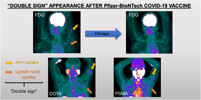
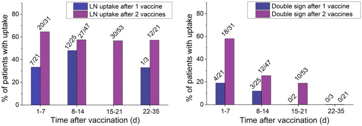
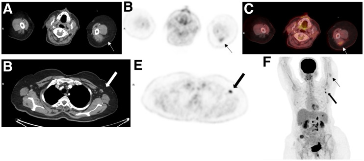
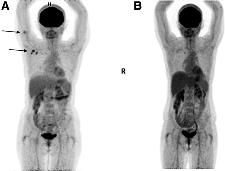
Vaccination against coronavirus 2019 (COVID-19) has created new challenges. Lymphadenopathy with increased uptake in patients undergoing PET/CT may mislead to unnecessary further evaluation. We have analyzed routinely performed PET/CT studies after Pfizer-BioNTech vaccination to familiarize ourselves with the PET/CT appearance of various PET tracers and to prevent the consequences of misinterpretation. Methods: We analyzed 1,018 PET/CT studies performed between January 1, 2021, and February 15, 2021. Information about the dates and sites of vaccination was collected. Visual and semiquantitative analysis of axillary-neck lymphadenopathy and arm uptake was correlated with immunization data. Results: Increased uptake in axillary lymphadenopathy was observed unilaterally in 66% of vaccinated patients, in 55% of patients vaccinated once, and in 69% of those vaccinated twice. The intensity of uptake decreased over time. Fifty-four of 274 patients (20%) had simultaneous increased activity in the posterior arm and ipsilateral axillary lymphadenopathy (double sign [DS]). The sensitivity, specificity, positive predictive value, and negative predictive value were 55.4%, 83.6%, 86.7%, 49.2%, respectively, for axillary lymphadenopathy and 38.6%, 100%, 100%, and 66.1%, respectively, for DS. No DS was observed later than 10 and 21 d after the first and the second vaccinations, respectively. None of the nonvaccinated patients had arm uptake or DS. Conclusion: Vaccination against COVID-19 frequently causes nonspecific axillary lymphadenopathy with increased PET tracer activity. In one fifth of our study population, this lymphadenopathy was associated with increased uptake at the vaccination site, DS. DS was 100% specific, with a 100% positive predictive value for postvaccination lymphadenopathy, hence enabling avoidance of misinterpretation of PET/CT studies and further unnecessary evaluation.
Keywords: COVID-19; PET/CT; lymphadenopathy; pattern; vaccination.
© 2022 by the Society of Nuclear Medicine and Molecular Imaging.
Figures
References
MeSH terms
Substances
LinkOut - more resources
Full Text Sources
Medical