Review
. 2021 Oct;62(10):1323-1329.
doi: 10.2967/jnumed.120.251512. Epub 2021 Jul 22.

Somatostatin Receptor Imaging and Theranostics: Current Practice and Future Prospects

Affiliations
Review

Somatostatin Receptor Imaging and Theranostics: Current Practice and Future Prospects

Sonya Park et al. J Nucl Med. 2021 Oct.

Abstract

A new era of precision diagnostics and therapy for patients with neuroendocrine neoplasms began with the approval of somatostatin receptor (SSTR) radiopharmaceuticals for PET imaging followed by peptide receptor radionuclide therapy (PRRT). With the transition from SSTR-based γ-scintigraphy to PET, the higher sensitivity of the latter raised questions regarding the direct application of the planar scintigraphy-based Krenning score for PRRT eligibility. Also, to date, the role of SSTR PET in response assessment and predicting outcome remains under evaluation. In this comprehensive review article, we discuss the current role of SSTR PET in all aspects of neuroendocrine neoplasms, including its relation to conventional imaging, selection of patients for PRRT, and the current understanding of SSTR PET-based response assessment. We also provide a standardized reporting template for SSTR PET with a brief discussion.

Keywords: 68Ga-DOTANOC; 68Ga-DOTATATE; SSTR; neuroendocrine neoplasms; peptide receptor radionuclide therapy; somatostatin.


Figures

FIGURE 1.
FIGURE 1.
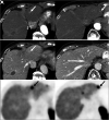
Pancreatic NEN with hepatic metastases (arrows). (A) Abdominal contrast-enhanced CT showing 2 small arterially enhancing left hepatic lesions. (B) Corresponding portal venous phase, where lesions are less conspicuous. (C) Transaxial PET images showing 68Ga-DOTATATE avidity in same lesions.
FIGURE 2.
FIGURE 2.
Abdominal contrast-enhanced MRI (with gadoxetate disodium) in patient with pancreatic NEN with hepatic metastases. (A and B) On arterial phase (A) and 20-min delayed-phase (B) images, 2 metastatic lesions (arrows) show arterial enhancement and contrast washout during delayed phase. (C and D) On arterial phase (C) and delayed-phase (D) images, size of previous lesions on 18-mo follow-up MRI has increased. Lesions are better delineated during delayed phase, facilitating accurate size measurements.
FIGURE 3.
FIGURE 3.
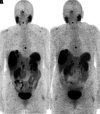
68Ga-DOTATATE (A) and 64Cu-DOTATATE (B) maximum-intensity-projection PET images of metastatic NEN showing similar findings. Both studies were performed as part of PET/MRI, with uptake times for 68Ga-DOTATATE and 64Cu-DOTATATE being 113 and 118 min, respectively (3 min/bed position for both).
FIGURE 4.
FIGURE 4.
Maximum-intensity projection images of patient with metastatic grade 1 (Ki-67 < 2%) NEN from small-bowel primary. (A) 68Ga-DOTATATE PET shows prominent uptake in primary tumor, lymphadenopathy, and liver metastases. (B) 18F-FDG PET shows no abnormal uptake (arrow points out incidentally noted fractured rib).
FIGURE 5.
FIGURE 5.
A 60-y-old woman with small-bowel NET on octreotide therapy. 68Ga-DOTATATE PET/CT (maximum-intensity projection [A], transaxial PET [B], CT [C], PET/CT [D]) shows prominent uptake at tumor sites. This finding would make patient eligible for PRRT, but overall limited extent of disease in liver and retroperitoneum favors surgical resection over PRRT.

References

    1. Cives M, Strosberg JR. Gastroenteropancreatic neuroendocrine tumors. CA Cancer J Clin. 2018;68:471–487. - PubMed
    1. Rindi G, Klimstra DS, Abedi-Ardekani B, et al. . A common classification framework for neuroendocrine neoplasms: an International Agency for Research on Cancer (IARC) and World Health Organization (WHO) expert consensus proposal. Mod Pathol. 2018;31:1770–1786. - PMC - PubMed
    1. Nagtegaal ID, Odze RD, Klimstra D, et al. . The 2019 WHO classification of tumours of the digestive system. Histopathology. 2020;76:182–188. - PMC - PubMed
    1. Krenning EP, Kwekkeboom DJ, Bakker WH, et al. . Somatostatin receptor scintigraphy with [111In-DTPA-d-Phe1]- and [123I-Tyr3]-octreotide: the Rotterdam experience with more than 1000 patients. Eur J Nucl Med. 1993;20:716–731. - PubMed
    1. Rufini V, Calcagni ML, Baum RP. Imaging of neuroendocrine tumors. Semin Nucl Med. 2006;36:228–247. - PubMed

MeSH terms

Substances