Review
. 2021 Jul;73(3):1118-1149.
doi: 10.1124/pharmrev.120.000097.

Therapeutic Targeting of α 7 Nicotinic Acetylcholine Receptors

Affiliations
Review

Therapeutic Targeting of α 7 Nicotinic Acetylcholine Receptors

Roger L Papke et al. Pharmacol Rev. 2021 Jul.

Abstract

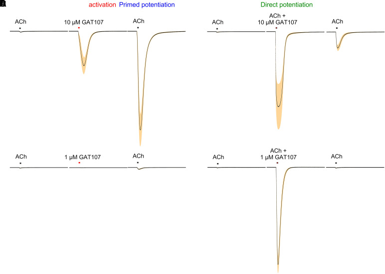
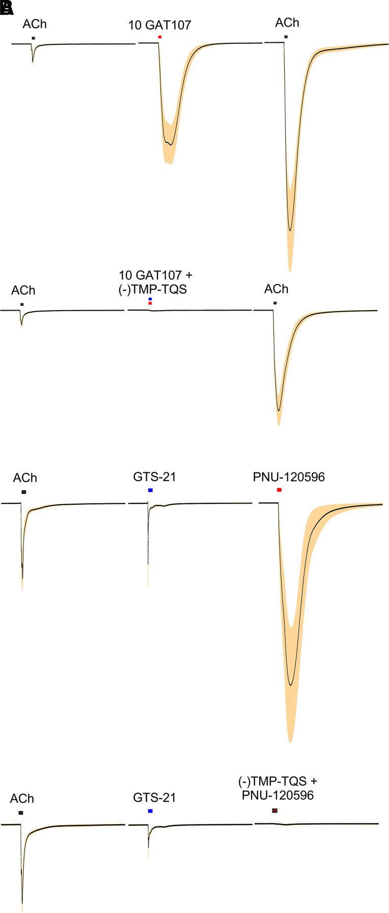
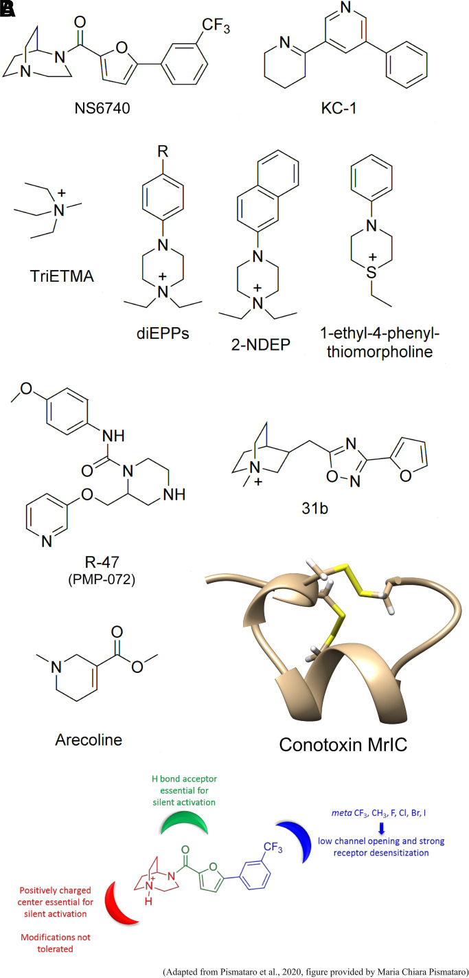
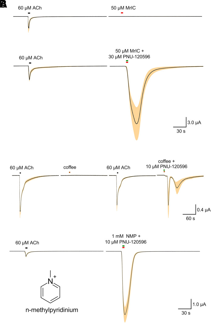
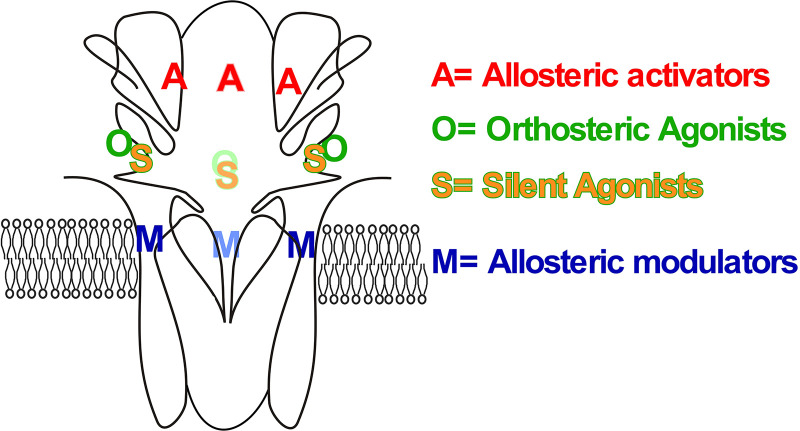
The α7-type nicotinic acetylcholine receptor is one of the most unique and interesting of all the members of the cys-loop superfamily of ligand-gated ion channels. Since it was first identified initially as a binding site for α-bungarotoxin in mammalian brain and later as a functional homomeric receptor with relatively high calcium permeability, it has been pursued as a potential therapeutic target for numerous indications, from Alzheimer disease to asthma. In this review, we discuss the history and state of the art for targeting α7 receptors, beginning with subtype-selective agonists and the basic pharmacophore for the selective activation of α7 receptors. A key feature of α7 receptors is their rapid desensitization by standard "orthosteric" agonist, and we discuss insights into the conformational landscape of α7 receptors that has been gained by the development of ligands binding to allosteric sites. Some of these sites are targeted by positive allosteric modulators that have a wide range of effects on the activation profile of the receptors. Other sites are targeted by direct allosteric agonist or antagonists. We include a perspective on the potential importance of α7 receptors for metabotropic as well as ionotropic signaling. We outline the challenges that exist for future development of drugs to target this important receptor and approaches that may be considered to address those challenges. SIGNIFICANCE STATEMENT: The α7-type nicotinic acetylcholine receptor (nAChR) is acknowledged as a potentially important therapeutic target with functional properties associated with both ionotropic and metabotropic signaling. The functional properties of α7 nAChR can be regulated in diverse ways with the variety of orthosteric and allosteric ligands described in this review.


Conflict of interest statement

No author has an actual or perceived conflict of interest with the contents of this article.

Figures

Fig. 1
Fig. 1
Compounds used to determine the structural motifs of α7-selective agonists as presented in (Horenstein et al. 2008). (A) α7-Selective agonists are in red boxes compared with related compounds that are not selective for α7. The highlighted compounds are tilorone (2,7-bis[2-(diethylamino)ethoxy]fluoren-9-one dihydrochloride) (Briggs et al.,2008); A-844606 (2-(5-methyl-hexahydro-pyrrolo[3,4-c]pyrrol-2-yl)-xanthen-9-one) (Briggs et al., 2008); ACME (cis-1-methyl-2,3,3a,4,5,9b,-hexahydro-1H-pyrrolo[3,2-h]isoquinoline) (Papke et al., 2005b); S 24795 (2-[2-(4-bromophenyl)-2-oxoethyl]-1-methyl pyridinium) (Lopez-Hernandez et al., 2007); tropane ((1R,5S)-8-methyl-8-azabicyclo[3.2.1]octane) (Papke et al., 2005a); tropinone (8-methyl-8-azabicyclo[3.2.1]octan-3-one) (Papke et al., 2005a); tropisetron ([(1R,5S)-8-methyl-8-azabicyclo[3.2.1]octan-3-yl] 1H-indole-3-carboxylate) (Macor et al., 2001; Papke et al., 2005a); cocaine methiodide, (methyl (1R,2R,3S,5S)-3-benzoyloxy-8,8-dimethyl-8-azoniabicyclo[3.2.1]octane-2-carboxylate) (Francis et al., 2001); AR-R17779 ((−)-Spiro-1-azabicyclo[2.2.2]octane-3,5′-oxazolidin-2'-one) (Mullen et al., 2000; Papke et al., 2004); JN403 ((S)-(1-azabicyclo[2.2.2]oct-3-yl)-carbamic acid (S)-1-(2-fluoro-phenyl)-ethyl ester) (Feuerbach et al., 2007); ABBF (N-[(3R)-1-azabicyclo[2.2.2]oct-3-yl]-7-[2-(methoxy)phenyl]-1-benzofuran-2-carboxamide) (Boess et al., 2007); PNU-282987 (Bodnar et al., 2005); PHA-543,613 (Acker et al., 2008); compound 15b ((+)-3-[2-(benzo[b]thiophen-2-yl)-2- oxoethyl]-1-azabicyclo[2.2.2]octane) (Tatsumi et al., 2004); compound 25 ((R)-3′-(5-chlorothiophen-2-yl)spiro-1-azabicyclo[2.2.2]octane-3,5′-[1',3′]oxazolidin-2'-one) (Tatsumi et al., 2004); compound 23 ((R)-3′-(5-iodothiophen-2-yl)spiro [1-azabicyclo[2.2.2]octane-3,5′-[1',3′]oxazolidin]-2'-one) (Tatsumi et al., 2004); PSAB-OFP, ((R)-(-)-5′phenylspiro[1-azabicyclo[2.2.2] octane-3,2'-(3′H)furo[2,3-b]pyridine) (Broad et al., 2002); SSR-180711 (1,4-diazabicyclo[3.2.2]nonane-4-carboxylic acid, 4-bromophenyl ester) (Biton et al., 2007); TC-1698 (2-(3-pyridyl)-1-azabicyclo[3.2.2]nonane) (Marrero et al., 2004); and PHA-709829 (Acker et al., 2008). (B) Modified quinuclidine α7-selective agonists: quinuclidinol (1-azabicyclo[2.2.2]octan-3-ol) (Horenstein et al., 2008); methyl-quinuclidine (1-methyl-1-azoniabicyclo[2.2.2]octane iodide) (Horenstein et al., 2008); and BQNE ((E)-3-benzylidene-1-azoniabicyclo-[2.2.2]octane chloride) (Horenstein et al., 2008).
Fig. 2
Fig. 2
Recently identified putative α7-selective agonists (see Table 1). A-582941 (2-methyl-5-[6-phenylpyridazin-3-yl]octa- hydropyrrolo[3,4-c]pyrrole) (Tietje et al., 2008); ABT-107, 5-(6-[(3R)-1-azabicyclo[2,2,2]oct-3-yloxy] pyridazin-3-yl)-1H-indole (Malysz et al., 2010); ABT-126, (4s)-4-(5-phenyl-1, 3, 4-thiadiazol-2-yloxy)-1-azatricyclo[3.3.1.13, 7]decane (Haig et al., 2018); AQW051, (R)-3-(6-p-tolyl-pyridin-3-yloxy)-1-aza-bicyclo (2.2.2)octane (Feuerbach et al., 2015); AZD0328, (20 R)-spiro-[1-azabicyclo[2.2.2]octane-3,20 (30 H)-furo[2,3-b]pyridine] d-tartrate (Sydserff et al., 2009); BMS-933043, (2R)-N-(6-(1H-imidazol-1-yl)-4-pyrimidinyl)-4′H-spiro[4-azabicyclo[2.2.2]octane-2,5′-[1,3]oxazol]-2′-amine (Cook et al., 2016; Pieschl et al., 2017); BMS-910731, N-(6-methyl-1,3-benzoxazol-2-yl)-3′,5′-dihydro-4-azaspiro[bicyclo[2.2.2]octane-2,4'-imidazole]-2'-amine (Hill et al., 2017); BMS-902483, (1S,2R,4S)-N-isoquinolin-3-yl)-4′H-4-azaspiro[bicyclo[2.2.2]octane-2,5′oxazol]-2′-amine (Hill et al., 2016; 28105289) (Cook et al., 2016); Br-IQ17B, N-[(3R)-1- azabicyclo[2,2,2]oct-3-yl]-5-bromoindolizine-2-carboxamide (Tang et al., 2015); CP-810,123, 4-(5-methyloxazolo[4,5-b]pyridin-2-yl)-1,4-diazabicyclo[3.2.2]nonane (O'Donnell et al., 2010); EVP-6124 (Prickaerts et al., 2012); FRM-17874 (Stoiljkovic et al., 2015); NS6784, 2-(1,4-diazabicyclo[3.2.2] nonan-4-yl)-5-phenyl-1,3,4-oxadiazole (Briggs et al., 2009); SEN12333, WAY-317538 5-morpholin-4-yl-pentanoic acid (4-pyridin-3-yl-phenyl)-amide (Roncarati et al., 2009); SEN15924, WAY-361789, 5-(4-acetyl[1,4]diazepan-1-yl)pentanoic acid [5-(4-methoxyphenyl)-1H-pyrazol-3-yl] amide (Zanaletti et al., 2012b); SEN78702, WYE-308775, N‐[5-(5-fluoropyridin-3-yl)‐1H‐pyrazol-3-yl]-4-piperidin-1-ylbutyramide (Zanaletti et al., 2012a); TC-7020, [5-methyl-N-[2-(pyridin- 3-ylmethyl)-1-azabicyclo[2.2.2]oct-3-yl]thiophene-2-carbox-amide (Marrero et al., 2010); and 5-(1-((1S,3R)-8-methyl-8-azabicyclo[3.2.1]octan-3-yl)-1H-1,2,3-triazol-4-yl)-1H-indole (TTIn-1) and related compounds (Arunrungvichian et al., 2015).
Fig. 3
Fig. 3
Apparent potentiation of α7 peak current responses by tonic low concentrations of agonist. (A) Averaged normalized responses (±S.E.M.) of oocytes (n = 7) expressing human α7 to repeated applications of ACh. After the first two control applications, the bath solution was switched to Ringer's solution containing 300 nM ACh. (B) Peak current response data of “representative” (i.e., n = 1) responses of an α7-expressing oocyte extrapolated from Figure 6B of (Prickaerts et al. 2012).
Fig. 4
Fig. 4
Structures of commonly used α7 PAMs. See Table 2 for chemical names and references. NS-1738 and N-(4-chlorophenyl)-α-[[(4-chloro-phenyl)amino]methylene]-3-methyl-5-isoxazoleacet-amide (CCMI) are classified as type I (see text and Figure 5), whereas PNU-120596, (±)TQS, and A-867744 are type II PAMs. PAM-2 activity is intermediate between the two types (see text). The scaffold of TQS has provided the basis for many different allosteric ligands with diverse functions (Gill et al., 2012; Gill-Thind et al., 2015), as discussed in the text.
Fig. 5
Fig. 5
Representative data from prototypical type I (NS-1738) and type II (PNU-120596) PAMs. (A) Averaged normalized responses (±S.E.M.) of oocytes (n = 4) expressing human α7 to 60 µM ACh or 60 µM ACh coapplied with 10 µM NS-1738. The insert shows the control and potentiated responses scaled to the same peak amplitude. (B) Averaged normalized responses (±S.E.M.) of oocytes (n = 4) expressing human α7 to 60 µM ACh or 60 µM ACh coapplied with 10 µM PNU-120596. The insert shows the control and potentiated responses scaled to the same peak amplitude.
Fig. 6
Fig. 6
Schematic illustration of energy landscapes for the conformational states associated with α7 activation, desensitization, and modulation [adapted from (Quadri et al. 2019) and (Williams et al. 2011b)] illustrating their relative energy levels and transition rates. Under equilibrium conditions, the distributions of receptors into the resting closed, open, and desensitized states will be determined by the relative free energy of the states (represented by vertical displacements). Dynamically, the transition rates between the states will be inversely related to the log of the energy barriers between the states. In the absence of any PAM (center row), the primary effect of agonist binding is to shift the equilibrium between the conformational states from the resting closed (C) state toward the desensitized states Ds and Di with a small probability of opening only at relatively low levels of agonist occupancy. The shallow energy well assigned to the open state (O*) is consistent with the brief opening observed in single-channel recordings and the high energy barriers into the O* state consistent with the low Popen observed. With the binding of a type I PAM (upper row), the primary effect is to deepen the well for the open state and to permit repeated transitions between the threshold activation (Flip) state and the open state, consistent with observations of single-channel currents in the presence of the type I PAM (Andersen et al., 2016). Note that this effect is only seen at low levels of agonist occupancy (in box). In the presence of the type II PAM (lower row, the Ds state is connected to another Flip state that then permits many reopenings to full and subconductance open states (O', O'', and O''').
Fig. 7
Fig. 7
TQS isomers. Shown on top are the structures of the two isomers of TQS (Stokes et al., 2019). The upper traces are averaged normalized responses (±S.E.M.) of oocytes expressing human α7 to 60 µM ACh or 60 µM ACh coapplied with 10 µM (+)TQS or (−)TQS (n equal to 3 and 4, respectively). The lower traces are averaged normalized responses (±S.E.M.) of oocytes (n = 8) expressing human α7 to 60 µM ACh or 60 µM ACh coapplied with 0.3 µM (+)TQS or (−)TQS. The data for the 0.3 µM responses have previously been published in bar graph format (Stokes et al., 2019).
Fig. 8
Fig. 8
Ago-PAM structures. GAT107 (Thakur et al., 2013), which is the active isomer of 4BP-TQS (Gill et al., 2011), and B-973B (Garai et al., 2018), which is the active isomer of B-973, 3-(3,4-difluorophenyl)-N-(1-(6-(4-(pyridin-2-yl)piperazin-1-yl)pyrazin-2-yl)ethyl)propanamide (Post-Munson et al., 2017).
Fig. 9
Fig. 9
Concentration and protocol dependence of responses to GAT107. (A) The traces are the averaged normalized responses (±S.E.M.) of oocytes expressing human α7 to 10 µM GAT107 applied alone and followed by an application of 60 µM, as compared with the initial responses to ACh alone (n = 5). (B) The traces shown are the averaged normalized responses (±S.E.M.) of oocytes expressing human α7 to 10 µM GAT107 coapplied with 60 µM ACh and followed by an application of 60 µM, as compared with the initial responses to ACh alone (n = 5). (C) The traces shown are the averaged normalized responses (±S.E.M.) of oocytes expressing human α7 to 1 µM GAT107 applied by itself and followed by an application of 60 µM, as compared with the initial responses to ACh alone (n = 7). (D) The traces shown are the averaged normalized responses (±S.E.M.) of oocytes expressing human α7 to 1 µM GAT107 coapplied with 60 µM ACh and followed by an application of 60 µM, as compared with the initial responses to ACh alone (n = 7).
Fig. 10
Fig. 10
Allosteric antagonism by (−)TMP-TQS. (A) The upper traces show the averaged normalized responses (±S.E.M.) of oocytes expressing human α7 to 10 µM GAT107 applied alone and followed by an application of 60 µM, as compared with the initial responses to ACh alone (n = 5) using the same protocol illustrated in Figure 9. The lower traces show the averaged normalized responses (±S.E.M.) obtained with the same protocol but with the coapplication of 100 µM (−)TQS with 10 µM GAT107 (n = 7). (B) Inhibition of responses to PNU-120596 after desensitization produced by 100 µM GTS-21. The upper traces show the averaged normalized responses (±S.E.M.) of oocytes (n = 4) expressing human α7 to a control application of ACh, an application of 100 µM GTS-21 and then the application of 10 µM PNU-120596 alone. The lower traces show the averaged normalized responses (±S.E.M.) obtained with the same protocol but with the coapplication of 100 µM (−)TQS with 10 µM PNU-120596 (n = 6).
Fig. 11
Fig. 11
Silent agonists. (A) Structures: NS6740, 1,4-diazabicyclo[3.2.2]non-4-yl[5-[3-(trifluoromethyl)phenyl]-2-furanyl]methanone hydrochloride (Pismataro et al., 2020); KC-1 (Chojnacka et al., 2013); TriETMA, triethylmethylammonium (Papke et al., 2014a); diEPP (Papke et al., 2014a); DMPP, 1,1-dimethyl-4-phenylpiperazin-1-ium iodide (Quadri et al., 2016); 2-NDEP, 1,1-diethyl-4(naphthalene-2-yl)piperazin-1-ium (Gulsevin et al., 2019); R-47 (PMP-072), (R)-N-(4-methoxyphenyl)-2-((pyridin-3-yloxy)methyl)piperazine-1-carboxamide (Clark et al., 2014); 31b, 3-(furan-2-yl)-5-(quinuclidin-3-ylmethyl)-1,2,4-oxadiazole methiodide (Quadri et al., 2018b); and a-conotoxin MrIC (image provided by Dr. Alican Gulsevin). (B) Model for NS6740 pharmacophore adapted from (Pismataro et al., 2020) (image provided by Dr. Maria Chiara Pismataro).
Fig. 12
Fig. 12
Silent agonists revealed by PNU-120596 applications. (A) In the upper traces are the averaged normalized responses (±S.E.M.) of oocytes (n = 3) expressing human α7 to a control application of 60 µM ACh followed by an application of 50 µM conotoxin MrIC (Alomone, Jerusalem Israel). In the lower traces are the averaged normalized responses (±S.E.M.) of oocytes (n = 7) expressing human α7 to a control application of 60 µM ACh followed by an application of 50 µM conotoxin MrIC (Alomone, Jerusalem, Israel) coapplied with 30 µM PNU-120596. (B) The upper traces show the averaged normalized responses (±S.E.M.) of oocytes (n = 5) expressing human α7 to a control application of 60 µM ACh followed by an application of 1 ml room-temperature coffee. Cells were then stimulated with ACh again prior to a coapplication of coffee with 10 µM PNU-120596. The lower traces show the averaged normalized responses (±S.E.M.) of oocytes (n = 7) expressing human α7 to a control application of 60 µM ACh followed by an application of 1 mM of the coffee alkaloid N-methylpyridinium (insert) coapplied with 10 µM PNU-120596.
Fig. 13
Fig. 13
Antagonist structures. ASS234, N-({5-[3-(1-benzyl-4-piperidinyl)propoxy]-1-methyl-1H-indol-2-yl}methyl)-N-methyl-2-propyn-1-amine (Criado et al., 2016); compound 38, 2-(1-benzylpiperidin-4-yl)ethan-1-amine (Criado et al., 2016); compound 7i, N-((1-benzylpiperidin-4-yl)methyl)-1-(2-chlorobenzyl)-N-methyl-1H-1,2,3-triazole-4-carboxamide (Peng et al., 2010); B10, 3-(4-bromophenyl)-8-methyl-1-oxa-2,4,8-triazaspiro[4.5]dec-2-ene (Zhang et al., 2020a); mecamylamine, N,2,3,3-tetramethylbicyclo[2.2.1]heptan-2-amine;hydrochloride; tkP3BzPB, 1,2,4,5-tetra-{5-[1-(3-benzyl)pyridinium]pent-1-yl}benzene tetrabromide; and MLA.
Fig. 14
Fig. 14
Binding sites for therapeutic ligands on α7 nAChRs. Cartoon of a cut-away view of an α7 receptor subunit complex (two subunits removed) and the approximate locations of the binding sites for the ligands discussed in this review. Located in the extracellular vestibule are putative binding sites (A) for allosteric ligands, such as ago-PAMs, allosteric agonists (2NDEP), and allosteric antagonists/inverse agonists, such as (−)TMP-TQS. Located at subunit interfaces on the outer surface of the extracellular domain are the binding sites (O) for ACh and other orthosteric agonists. These sites will also overlap the sites (S) that bind somewhat larger silent agonists. The binding site for PAMs and other allosteric modulators (M) is within the transmembrane domain and requires specific residues at the outer end of the second transmembrane domain. This figure is adapted from (Papke and Lindstrom 2020).

References

    1. Abbas M, Alzarea S, Papke RL, Rahman S (2017) The α7 nicotinic acetylcholine receptor positive allosteric modulator attenuates lipopolysaccharide-induced activation of hippocampal IκB and CD11b gene expression in mice. Drug Discov Ther 11:206–211. - PMC - PubMed
    1. Acker BAJacobsen EJRogers BNWishka DGReitz SCPiotrowski DWMyers JKWolfe MLGroppi VEThornburgh BA, et al. (2008) Discovery of N-[(3R,5R)-1-azabicyclo[3.2.1]oct-3-yl]furo[2,3-c]pyridine-5-carboxamide as an agonist of the alpha7 nicotinic acetylcholine receptor: in vitro and in vivo activity. Bioorg Med Chem Lett 18:3611–3615. - PubMed
    1. Adams DJ, Nutter TJ (1992) Calcium permeability and modulation of nicotinic acetylcholine receptor-channels in rat parasympathetic neurons. J Physiol Paris 86:67–76. - PubMed
    1. Aiyar VN, Benn MH, Hanna T, Jacyno J, Roth SH, Wilkens JL (1979) The principal toxin of Delphinium brownii Rydb., and its mode of action. Experientia 35:1367–1368. - PubMed
    1. Alkondon M, Albuquerque EX (1995) Diversity of nicotinic acetylcholine receptors in rat hippocampal neurons. III. Agonist actions of the novel alkaloid epibatidine and analysis of type II current. J Pharmacol Exp Ther 274:771–782. - PubMed

Publication types