. 2021 Jul 23;11(1):15141.
doi: 10.1038/s41598-021-94565-y.

The role of IL-1 in adipose browning and muscle wasting in CKD-associated cachexia

Affiliations

The role of IL-1 in adipose browning and muscle wasting in CKD-associated cachexia

Wai W Cheung et al. Sci Rep. .

Abstract

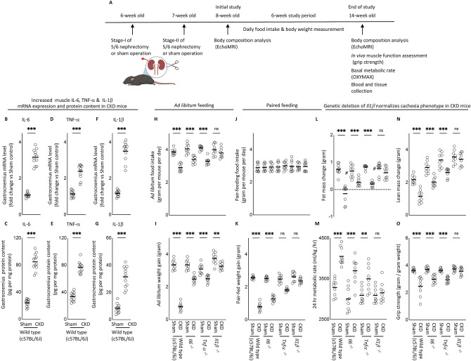
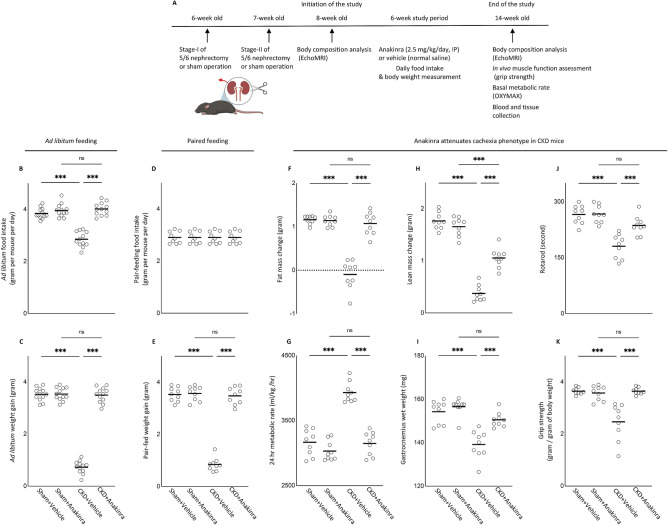
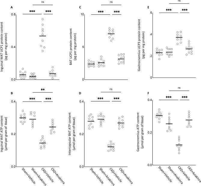
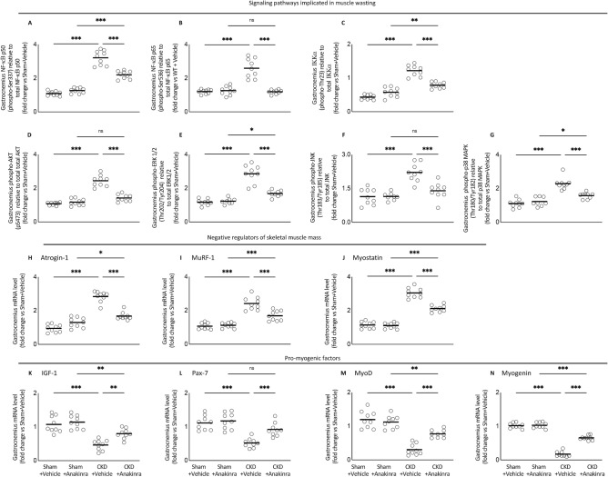
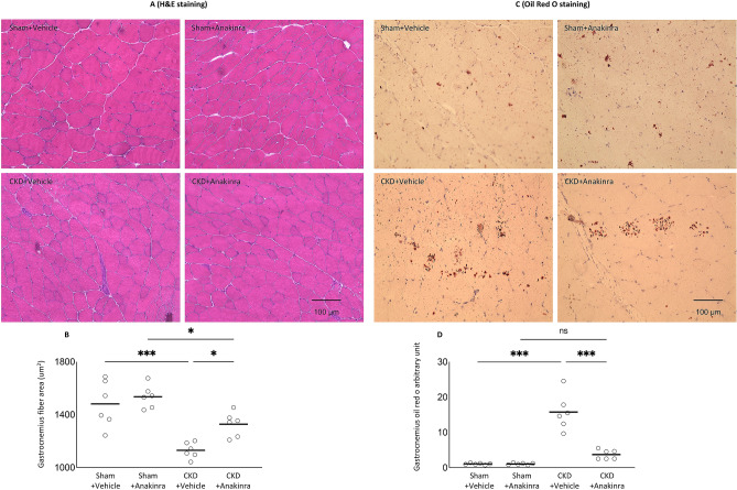
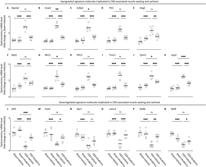
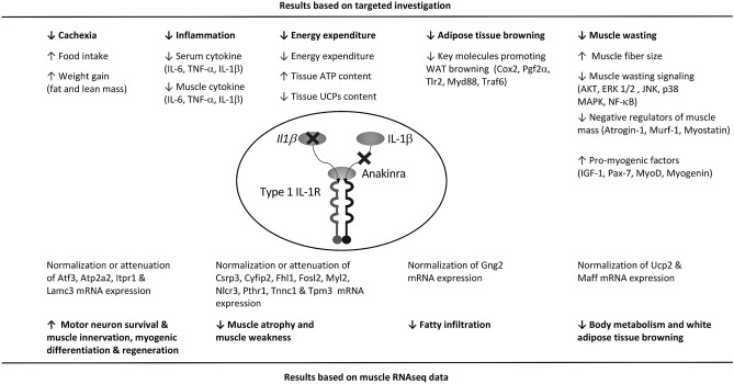
Cytokines such as IL-6, TNF-α and IL-1β trigger inflammatory cascades which may play a role in the pathogenesis of chronic kidney disease (CKD)-associated cachexia. CKD was induced by 5/6 nephrectomy in mice. We studied energy homeostasis in Il1β-/-/CKD, Il6-/-/CKD and Tnfα-/-/CKD mice and compared with wild type (WT)/CKD controls. Parameters of cachexia phenotype were completely normalized in Il1β-/-/CKD mice but were only partially rescued in Il6-/-/CKD and Tnfα-/-/CKD mice. We tested the effects of anakinra, an IL-1 receptor antagonist, on CKD-associated cachexia. WT/CKD mice were treated with anakinra (2.5 mg/kg/day, IP) or saline for 6 weeks and compared with WT/Sham controls. Anakinra normalized food intake and weight gain, fat and lean mass content, metabolic rate and muscle function, and also attenuated molecular perturbations of energy homeostasis in adipose tissue and muscle in WT/CKD mice. Anakinra decreased serum and muscle expression of IL-6, TNF-α and IL-1β in WT/CKD mice. Anakinra attenuated browning of white adipose tissue in WT/CKD mice. Moreover, anakinra normalized gastrocnemius weight and fiber size as well as attenuated muscle fat infiltration in WT/CKD mice. This was accompanied by correcting the increased muscle wasting signaling pathways while promoting the decreased myogenesis process in gastrocnemius of WT/CKD mice. We performed qPCR analysis for the top 20 differentially expressed muscle genes previously identified via RNAseq analysis in WT/CKD mice versus controls. Importantly, 17 differentially expressed muscle genes were attenuated in anakinra treated WT/CKD mice. In conclusion, IL-1 receptor antagonism may represent a novel targeted treatment for adipose tissue browning and muscle wasting in CKD.


Conflict of interest statement

The authors declare that there is no competing conflict of interest, either financial or non-financial as listed in the journal website, that could be perceived as prejudicing the impartiality of the research reported.

Figures

Figure 1
Figure 1
Increased muscle mRNA and protein content of IL-6, TNF-α and IL-1β in CKD mice and genetic depletion of Il1β provides a better rescue of cachexia in CKD mice compared to Il6 and Tnfα deficiency. Schematic experimental design is shown (A). (A) Was created with BioRender.com. Results of three experiments were shown. For the first experiment, CKD was induced by 5/6 nephrectomy in WT mice and sham operation was performed in WT control mice. Gene expression and protein content of pro-inflammatory cytokine IL-6, TNF-α and IL-1β in gastrocnemius muscle in WT/CKD and WT/Sham mice was performed. Data are expressed as mean ± SEM and results of WT/CKD mice were compared to WT/Sham mice (BG). For the second experiment, four groups of mice were included: WT/CKD and WT/Sham, Il6−/−/CKD and Il6−/−/Sham, Tnfα−/−/CKD and Tnfα−/−/Sham, Ilβ−/−/CKD and Ilβ−/−/Sham. All mice were fed ad libitum and food intake as well as weight gain in mice were recorded (H,I). For the third experiment, we investigated the metabolic benefits of genetic depletion of Il6, Tnfα and Il1β in CKD mice beyond the nutritional stimulation by employed a pair-feeding strategy. Four groups of mice are WT/CKD and WT/Sham, Il6−/−/CKD and Il6−/−/Sham, Tnfα−/−/CKD and Tnfα−/−/Sham, Ilβ−/−/CKD and Ilβ−/−/Sham. WT/CKD mice were fed ad libitum whereas other groups of mice were pair-fed to that of WT/CKD mice (J). Weight gain, fat and lean content, 24-h oxygen consumption and in vivo muscle function (grip strength) was measured in mice (KO). For the second and third experiments, data are expressed as mean ± SEM and results of WT/CKD, Il6−/−/CKD, Tnfα−/−/CKD and Il1β−/−/CKD mice were compared to WT/Sham, Il6−/−/Sham, Tnfα−/−/Sham and Il1β−/−/Sham, respectively.
Figure 2
Figure 2
Anakinra attenuates cachexia in CKD mice. WT/CKD and WT/Sham mice were treated with anakinra (2.5 mg/kg/day, IP, daily) or normal saline as a vehicle control for 6 weeks. Schematic experimental design is shown (A). Figure 2A was created with BioRender.com. Results Mice were fed ad libitum and food intake and weight gain in mice were recorded (B,C). To assess the beneficial effects of anakinra beyond its nutritional effects, we employed a pair-feeding strategy. WT/CKD mice treated with vehicle were given an ad libitum amount of food whereas other groups of mice were given an equivalent amount of food (D). Weight change, fat and lean mass content, 24-h oxygen consumption and in vivo muscle function (grip strength) was measured in mice (EK). Data are expressed as mean ± SEM. Results of WT/CKD + Vehicle were compared to WT/Sham + Vehicle and WT/CKD + Anakinra were compared to WT/Sham + Anakinra, respectively. In addition, results of WT/CKD + Anakinra were also compared to WT/CKD + Vehicle. * p < 0.05, ** p < 0.01, *** p < 0.001.
Figure 3
Figure 3
Anakinra attenuates serum and muscle expression of IL-6, TNF-α and IL-1β in CKD mice. At the end of 6 weeks of anakinra or vehicle treatment, WT/CKD and WT/Sham mice were sacrificed and serum cytokine concentration (AC) as well as gastrocnemius muscle mRNA levels (DF) and protein content (GI) of IL-6, TNF-α and IL-1β were measured. Data are expressed as mean ± SEM. Results of WT/CKD + Vehicle were compared to WT/Sham + Vehicle and WT/CKD + Anakinra were compared to WT/Sham + Anakinra, respectively. In addition, results of WT/CKD + Anakinra were also compared to WT/CKD + Vehicle. * p < 0.05, ** p < 0.01, *** p < 0.001.
Figure 4
Figure 4
Anakinra ameliorates energy homeostasis in skeletal and adipose tissue in CKD mice. UCP content (A,C,E) and ATP protein content (B,D,F) in adipose tissue (inguinal white adipose tissue and brown adipose tissue) and gastrocnemius muscle were measured. Final results were expressed in arbitrary units, with one unit being the mean level in WT/Sham + Vehicle mice. Results of WT/CKD + Vehicle were compared to WT/Sham + Vehicle and WT/CKD + Anakinra were compared to WT/Sham + Anakinra, respectively. In addition, results of WT/CKD + Anakinra were also compared to WT/CKD + Vehicle. * p < 0.05, ** p < 0.01, *** p < 0.001.
Figure 5
Figure 5
Anakinra attenuates adipose tissue browning in CKD mice. Protein content of beige adipocyte markers (CD137, Tmem26 and Tbx-1) in inguinal white adipose tissue was measured (AC). In addition, protein content of Cox2 signaling pathway (Cox2 and Pgf2α) and toll like receptor pathway (Tlr2, MyD88 and Traf6) in inguinal white adipose tissue was measured (DH). Results of WT/CKD + Vehicle were compared to WT/Sham + Vehicle and WT/CKD + Anakinra were compared to WT/Sham + Anakinra, respectively. In addition, results of WT/CKD + Anakinra were also compared to WT/CKD + Vehicle. * p < 0.05, ** p < 0.01, *** p < 0.001.
Figure 6
Figure 6
Anakinra attenuates signaling pathways implicated in muscle wasting in CKD mice. Gastrocnemius muscle relative phosphorylated NF-κB p50 (Ser337) / total p50 ratio (A), NF-κB p65 (Ser536) / total p65 ratio (B) and Iκκα (Thr23) / total Iκκα ratio (C) as well as muscle relative phospho-Akt (pS473) / total Akt ratio (D), ERK 1/2 (Thr202/Tyr204) / total ERK 1/2 ratio (E), JNK (Thr183/Tyr185) / total JNK ratio (F), p38 MAPK (Thr180/Tyr182) / total p38 MAPK ratio (G) in mice. In addition, gastrocnemius muscle expression of interested genes in mice was measured by qPCR. Transcriptional expression of negative regulators of skeletal muscle mass (Atrogin-1, MuRF-1 and Myostatin) (HJ) and pro-myogenic factors (IGF-1, Pax-7, MyoD and Myogenin) (KN) were expressed in arbitrary units, with one unit being the mean level in WT/Sham + Vehicle mice. Results of WT/CKD + Vehicle were compared to WT/Sham + Vehicle and WT/CKD + Anakinra were compared to WT/Sham + Anakinra, respectively. In addition, results of WT/CKD + Anakinra were also compared to WT/CKD + Vehicle. * p < 0.05, ** p < 0.01, *** p < 0.001.
Figure 7
Figure 7
Anakinra normalizes muscle fiber size and attenuates muscle fat infiltration in CKD mice. Representative photomicrographs of gastrocnemius with H&E staining (A). Average gastrocnemius cross-sectional area was measured (B). Visualization of quantification of fatty infiltration by Oil Red O analysis in gastrocnemius muscle (C,D). Final results were expressed in arbitrary units, with one unit being the mean staining intensity in vehicle-treated WT/Sham mice. Difference among various groups of mice were analyzed as in Fig. 2.
Figure 8
Figure 8
Anakinra attenuates expression of the top 17 differentiated expression gastrocnemius muscle genes in CKD mice. Gastrocnemius muscle expression of interested genes in mice was measured by qPCR. Transcriptional expression of upregulated signature molecules implicated in CKD-associated muscle wasting and cachexia (Atp2a2, Csrp3, Cyfip2, Fhl1, Gng2, Myl2, Nlrc3, Pth1r, Tncc1, Tpm3 and Ucp2) (AK) and downregulated signature molecules implicated in CKD-associated muscle wasting and cachexia (Atf3, Fosl2, Itpr1, Lamc3, Mafb and Maff) (LQ) were expressed in arbitrary units, with one unit being the mean level in WT/Sham + Vehicle mice. Results of WT/CKD + Vehicle were compared to WT/Sham + Vehicle and WT/CKD + Anakinra were compared to WT/Sham + Anakinra, respectively. In addition, results of WT/CKD + Anakinra were also compared to WT/CKD + Vehicle. * p < 0.05, ** p < 0.01, *** p < 0.001.
Figure 9
Figure 9
Summary of the beneficial effects of anakinra on cachexia, energy homeostasis, muscle wasting and adipose tissue browning in CKD mice.

Similar articles

Cited by

References

    1. Cohen S, Nathan JA, Goldberg AL. Muscle wasting in disease: Molecular mechanisms and promising therapies. Nat. Rev. Drug Discov. 2015;14:58–74. doi: 10.1038/nrd4467. - DOI - PubMed
    1. Furrer R, Handschin C. Muscle wasting diseases: Novel targets and treatments. Annu. Rev. Pharmacol. Toxicol. 2019;59:315–339. doi: 10.1146/annurev-pharmtox-010818-021041. - DOI - PMC - PubMed
    1. Deger SM, et al. Systemic inflammation is associated with exaggerated skeletal muscle protein catabolism in maintenance hemodialysis patients. JCI Insight. 2017;2:95185. doi: 10.1172/jci.insight.95185. - DOI - PMC - PubMed
    1. Londhe P, Guttridge DC. Inflammation induced loss of skeletal muscle. Bone. 2015;80:131–142. doi: 10.1016/j.bone.2015.03.015. - DOI - PMC - PubMed
    1. Zhou J, Liu B, Liang C, Li Y, Song YH. Cytokine signaling in skeletal muscle wasting. Trends Endocrinol. Metab. 2016;27:335–347. doi: 10.1016/j.tem.2016.03.002. - DOI - PubMed

Publication types