. 2021 Jul 23;11(1):15075.
doi: 10.1038/s41598-021-94566-x.

Development of thymic tumor in [LSL:KrasG12D; Pdx1-CRE] mice, an adverse effect associated with accelerated pancreatic carcinogenesis

Affiliations

Development of thymic tumor in [LSL:KrasG12D; Pdx1-CRE] mice, an adverse effect associated with accelerated pancreatic carcinogenesis

Sophie Liot et al. Sci Rep. .

Abstract

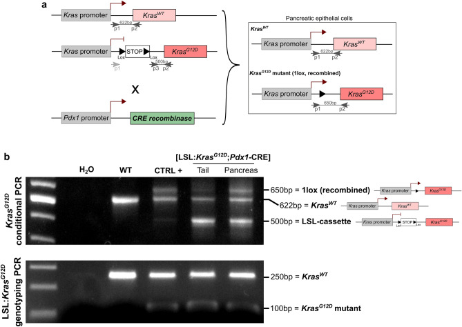
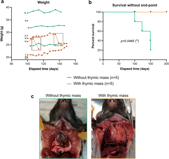
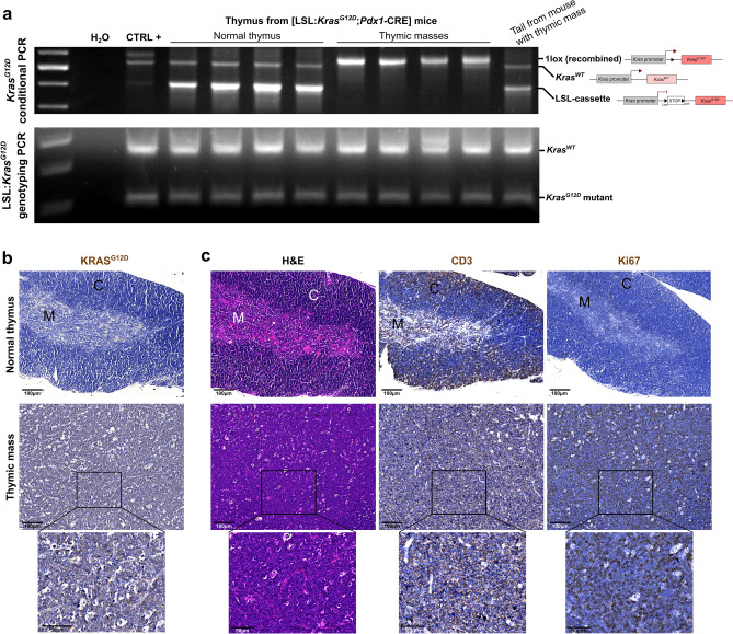
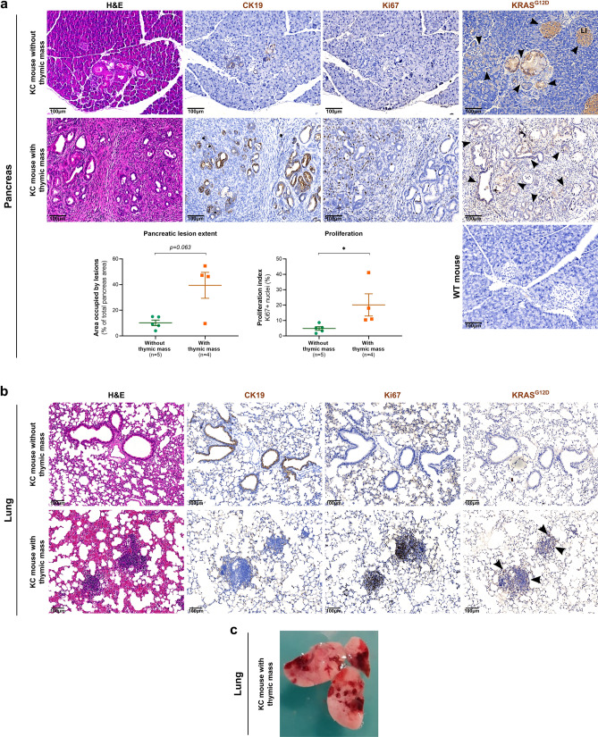
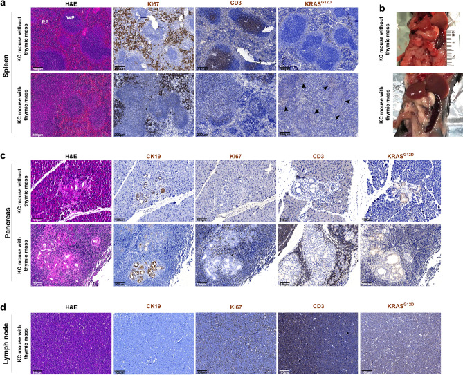
Pancreatic Ductal AdenoCarcinoma (PDAC) represents about 90% of pancreatic cancers. It is one of the most aggressive cancer, with a 5-year survival rate below 10% due to late diagnosis and poor therapeutic efficiency. This bad prognosis thus encourages intense research in order to better understand PDAC pathogenesis and molecular basis leading to the development of innovative therapeutic strategies. This research frequently involves the KC (LSL:KrasG12D;Pdx1-CRE) genetically engineered mouse model, which leads to pancreatic cancer predisposition. However, as frequently encountered in animal models, the KC mouse model also exhibits biases. Herein, we report a new adverse effect of KrasG12D mutation in KC mouse model. In our hands, 10% of KC mice developed clinical signs reaching pre-defined end-points between 100- and 150-days post-parturition, and associated with large thymic mass development. Histological and genetic analyses of this massive thymus enabled us (1) to characterize it as a highly proliferative thymic lymphoma and (2) to detect the unexpected recombination of the Lox-STOP-Lox cassette upstream KrasG12D allele and subsequent KRASG12D protein expression in all cells composing thymic masses. Finally, we highlighted that development of such thymic tumor was associated with accelerated pancreatic carcinogenesis, immune compartment disorganization, and in some cases, lung malignancies.


Conflict of interest statement

The authors declare no competing interests.

Figures

Figure 1
Figure 1
KC (LSL:KrasG12D:Pdx1-CRE) mouse model. (a) Schematic representation of the genetic construct for conditional KrasG12D expression under the control of Pdx1 promoter (CRE-Lox system). Primers used for KrasG12D conditional PCR are noticed. (b) Top: KrasG12D conditional PCR showing LSL cassette recombination in pancreas, but not tail, from KC mouse, and not in WT mouse. Bottom: KrasG12D genotyping PCR showing heterozygosity in KC mice (WT and KrasG12D alleles) and homozygosity in WT mouse (WT allele). Uncropped gels are provided in Supplementary Figure S2.
Figure 2:
Figure 2:
10% of KC mice exhibited rapid weight loss and decreased survival associated with thymic mass development. (a) Body weight monitoring of the KC mice presented in this study. Sex of the different mice is indicated on the left (M = male, F = female) (b) Cumulative survival proportion of the two groups presenting (orange, n = 5) or not (green, n = 5) a thymic mass, estimated using the Kaplan–Meier method. The two groups were compared thanks to Log-Rank (Mantel-Cox) statistical test. (c) Photo of the rib cage at autopsy, showing normal thymus (left) and the thymic mass developed in 10% of KC mice (right). Dotted line delimits thymus. Scale bars are indicated on the pictures.
Figure 3
Figure 3
Thymic mass showed a lymphoma-like histology and total LSL cassette recombination associated with KRAS G12D expression in all cells. (a) Top: KrasG12D conditional PCR showing LSL cassette recombination in thymic mass but not in normal thymus and tail (KC mice). Bottom: KrasG12D genotyping PCR showing heterozygosity in all KC mice of this study (WT and KrasG12D alleles). Uncropped gels are provided in Supplementary Figure S2. (b) Representative images of normal thymus and thymic mass immunolabelled for KRASG12D. (c) Representative images of normal thymus and thymic mass histologically analyzed (H&E staining and CD3e and Ki67 immunohistochemical labelings). C = cortex, M = medulla. Scale bars are indicated on the pictures.
Figure 4
Figure 4
KC mice developing thymic tumor exhibited accelerated pancreatic carcinogenesis, immune perturbations and lung malignancies. (a) Representative images of pancreas from KC mice showing or not thymic mass analyzed by classical histology (H&E staining) and immunohistochemistry (Ki67, CK19, and KRASG12D). Quantification of the area occupied by lesions in pancreas sections and of proliferating cells (Ki67-positive nuclei) are presented below the pictures. Groups were compared thanks to Mann–Whitney statistical test (non-parametric t test). (b) Representative images of histological analysis of lung from KC mice with or without thymic mass (H&E staining and Ki67, CK19 and KRASG12D immunohistochemistry). Bottom: This mouse displayed moderate lesions in lung. (c) Picture showing severe lung lesions, macroscopically visible, from KC mouse presenting thymic mass at autopsy. (a,b,d) Arrowheads highlight KRASG12D staining. Scale bars are indicated on the pictures.
Figure 5
Figure 5
Immune dysregulation in mice suffering from thymic lymphoma development. (a) Representative images of histological analysis of spleen from KC mice presenting or not thymic mass (H&E staining and Ki67 and KRASG12D immunohistochemistry). (b) Image showing enlarged spleen from KC mouse with thymic mass. (c) Representative images of histological analysis of the pancreas of KC mouse without thymic lymphoma, and one of the two mice with thymic mass which presented high level of immune infiltration (H&E staining and CK19, Ki67, CD3e and KRASG12D immunolabelings). (d) Representative images of histological analysis of the proximal lymph node of one of the two mice with thymic mass which presented high level of immune infiltration (H&E staining and CK19, Ki67, CD3e and KRASG12D immunolabelings). Scale bars are indicated on the pictures.

References

    1. Ferlay J, et al. Estimating the global cancer incidence and mortality in 2018: GLOBOCAN sources and methods. Int. J. Cancer. 2019;144:1941–1953. doi: 10.1002/ijc.31937. - DOI - PubMed
    1. Rahib L, et al. Projecting cancer incidence and deaths to 2030: The unexpected burden of thyroid, liver, and pancreas cancers in the United States. Cancer Res. 2014;74:2913–2921. doi: 10.1158/0008-5472.CAN-14-0155. - DOI - PubMed
    1. Quante AS, et al. Projections of cancer incidence and cancer-related deaths in Germany by 2020 and 2030. Cancer Med. 2016;5:2649–2656. doi: 10.1002/cam4.767. - DOI - PMC - PubMed
    1. Rawla P, Sunkara T, Gaduputi V. Epidemiology of pancreatic cancer: Global trends, etiology and risk factors. World J Oncol. 2019;10:10–27. doi: 10.14740/wjon1166. - DOI - PMC - PubMed
    1. Eibl, A. S. and G. Pancreatic Ductal Adenocarcinoma. Pancreapedia: The Exocrine Pancreas Knowledge Base (2015). 10.3998/panc.2015.14.

Publication types

MeSH terms

Substances

Supplementary concepts