Review
. 2021 Nov;22(11):733-750.
doi: 10.1038/s41580-021-00392-4. Epub 2021 Jul 23.

Machinery, regulation and pathophysiological implications of autophagosome maturation

Affiliations
Review

Machinery, regulation and pathophysiological implications of autophagosome maturation

Yan G Zhao et al. Nat Rev Mol Cell Biol. 2021 Nov.

Abstract

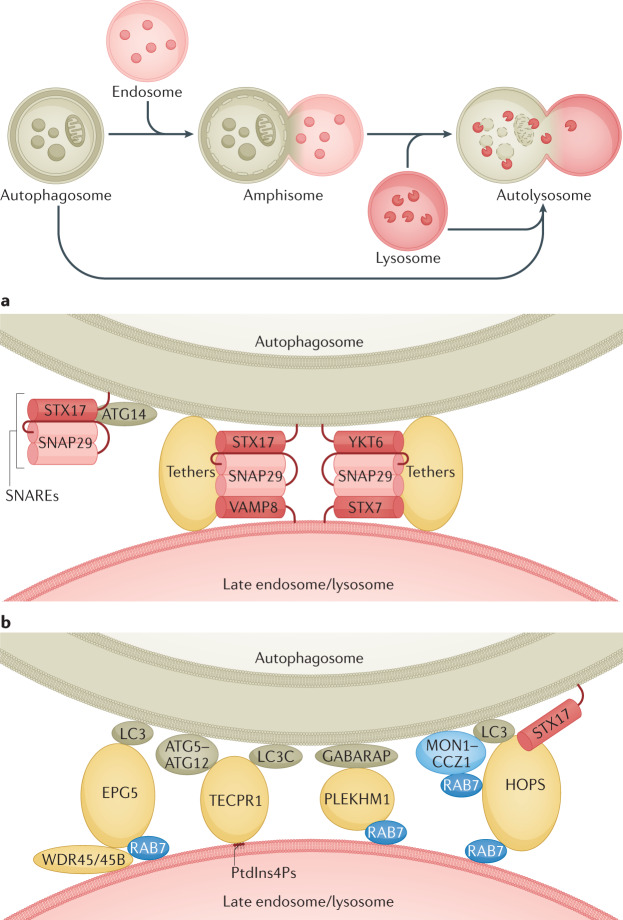
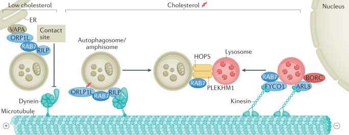
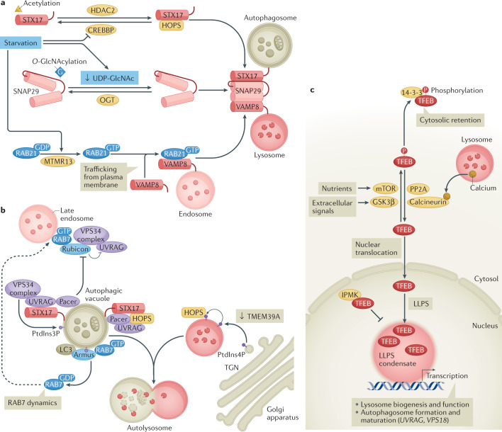
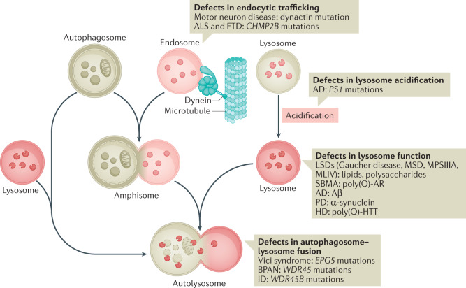
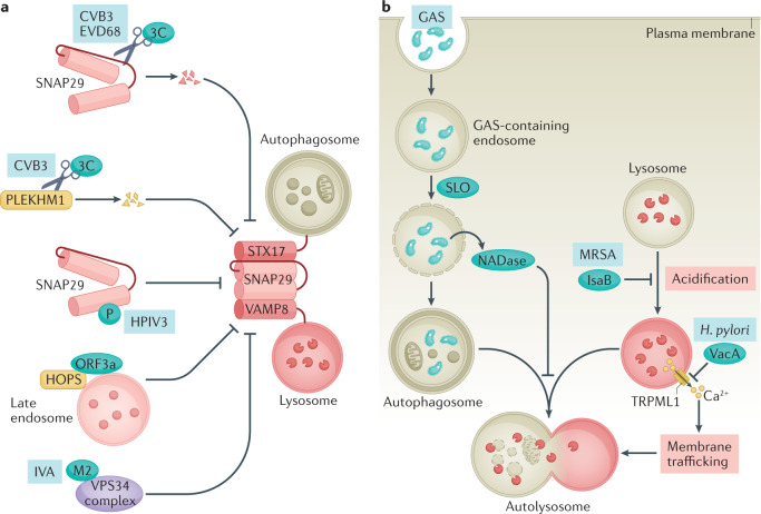
Autophagy is a versatile degradation system for maintaining cellular homeostasis whereby cytosolic materials are sequestered in a double-membrane autophagosome and subsequently delivered to lysosomes, where they are broken down. In multicellular organisms, newly formed autophagosomes undergo a process called 'maturation', in which they fuse with vesicles originating from endolysosomal compartments, including early/late endosomes and lysosomes, to form amphisomes, which eventually become degradative autolysosomes. This fusion process requires the concerted actions of multiple regulators of membrane dynamics, including SNAREs, tethering proteins and RAB GTPases, and also transport of autophagosomes and late endosomes/lysosomes towards each other. Multiple mechanisms modulate autophagosome maturation, including post-translational modification of key components, spatial distribution of phosphoinositide lipid species on membranes, RAB protein dynamics, and biogenesis and function of lysosomes. Nutrient status and various stresses integrate into the autophagosome maturation machinery to coordinate the progression of autophagic flux. Impaired autophagosome maturation is linked to the pathogenesis of various human diseases, including neurodegenerative disorders, cancer and myopathies. Furthermore, invading pathogens exploit various strategies to block autophagosome maturation, thus evading destruction and even subverting autophagic vacuoles (autophagosomes, amphisomes and autolysosomes) for survival, growth and/or release. Here, we discuss the recent progress in our understanding of the machinery and regulation of autophagosome maturation, the relevance of these mechanisms to human pathophysiology and how they are harnessed by pathogens for their benefit. We also provide perspectives on targeting autophagosome maturation therapeutically.


Conflict of interest statement

The authors declare no competing interests.

Figures

Fig. 1
Fig. 1. SNAREs, tethers and RAB proteins mediate autophagosome maturation.
a | Fusion between autophagosomes and late endosomes/lysosomes is mediated by two sets of SNARE complexes: the autophagosomal Qa SNARE STX17, the Qbc SNARE SNAP29 and the endolysosomal R SNARE VAMP8 (centre left); and the autophagosomal R SNARE YKT6, SNAP29 and the endolysosomal Qa SNARE STX7 (centre right). The unique hairpin structure formed by the two transmembrane domains of STX17 and the amino-terminal longin domain of YKT6 are involved in their autophagosomal recruitment,. The autophagosome-associated autophagy protein ATG14 interacts with STX17 to promote the assembly of the STX17–SNAP29 subcomplex (left). The assembly and function of both complexes are facilitated by tether proteins. b | Multiple tether proteins are involved in fusion of autophagosomes with late endosomes/lysosomes. EPG5 is targeted to autophagosomes and lysosomes by binding to LC3 and RAB7, respectively. In neural cells, WDR45 and WDR45B facilitate the lysosomal localization of EPG5. TECPR1 interacts with LC3C and ATG5–ATG12 on autophagosomes and phosphatidylinositol 4-phosphate (PtdIns4P) on lysosomes. RAB7 binds to the HOPS complex and PLEKHM1. On the autophagosome side, the HOPS complex binds to STX17 and LC3 proteins, and PLEKHM1 binds to GABARAPs. The active GTP-bound RAB7 associates with the membrane. Localization of RAB7 on autophagic vacuoles is facilitated by the LC3-binding MON1–CCZ1 guanine nucleotide exchange factor complex,.
Fig. 2
Fig. 2. Coupling transport and fusion of autophagic vacuoles and late endosomes/lysosomes.
Autophagosomes, which form throughout the cytoplasm, and lysosomes, which are localized mainly in the perinuclear region, undergo bidirectional movement on microtubules. The late endosome/lysosome-localized BORC–ARL8 complex and the FYCO1–RAB7 pair recruit kinesin motors for anterograde transport of late endosomes/lysosomes. The RAB7 effectors RILP and ORP1L promote the membrane association of the dynein–dynactin motor machinery to mediate the retrograde transport of autophagic vacuoles (as well as late endosomes/lysosomes (not shown)). When cholesterol levels are low, ORP1L interacts with the endoplasmic reticulum (ER) protein VAPA to form ER–autophagosome membrane contacts, which releases dynein–dynactin and prevents retrograde trafficking, thereby interfering with autophagosome maturation. During transport, the tethering factors HOPS complex and PLEKHM1 are concomitantly recruited to promote fusion processes (see Fig. 1).
Fig. 3
Fig. 3. Multiple mechanisms regulate autophagosome maturation.
a | The SNARE domain of STX17 is modified by acetylation, a process controlled by the acetyltransferase CREBBP and the deacetylase HDAC2. Starvation inactivates CREBBP, resulting in deacetylation of STX17. Deacetylated STX17 interacts more strongly with the HOPS complex and SNAP29 and thus promotes autophagosome–lysosome fusion. SNAP29 is O-GlcNAcylated by O-linked β-N-acetylglucosamine (O-GlcNAc) transferase (OGT). This modification attenuates the assembly of SNAP29-containing SNARE complexes. Under starvation conditions that decrease the intracellular UDP-GlcNAc level, or in OGT-knockdown cells, the O-GlcNAcylation of SNAP29 is reduced, which in turn facilitates the assembly of the trans-SNARE complex for autophagosome maturation. MTMR13, the guanine nucleotide exchange factor for the endosomal protein RAB21, controls RAB21-dependent trafficking of plasma membrane-localized VAMP8 to late endosomes/lysosomes. Upon starvation, MTMR13 activates RAB21, which subsequently promotes the translocation of VAMP8 R SNARE to late endosomes/lysosomes to promote fusion of endosomes/lysosomes with autophagic vacuoles (see Fig. 1). b | Phosphatidylinositol 3-phosphate (PtdIns3P) on autophagic vacuoles, generated by the UVRAG-containing VPS34 complex, facilitates autophagosome maturation by recruiting the tethering factors HOPS complex. Rubicon interacts with the UVRAG–VPS34 complex and negatively regulates its function. Pacer is targeted by autophagic vacuole-localized SNARE STX17 and phosphoinositides (PtdIns3P), and it antagonizes Rubicon and recruits the UVRAG–VPS34 complex to autophagic vacuoles. Pacer and UVRAG also recruit the HOPS complex. The RAB7 GTPase-activating protein Armus is targeted to autophagic vacuoles by interacting with LC3 and PtdIns3P, and promotes RAB7 dynamics, whereby it is recycled from the autophagic vacuole membranes. This generates a mobile pool of RAB7 that can be recruited to endosomes to drive their maturation to late endosomes/lysosomes. Depletion of the endoplasmic reticulum-localized transmembrane protein TMEM39A increases phosphatidylinositol 4-phosphate (PtdIns4P) levels on late endosomes/lysosomes (via inhibition of endoplasmic reticulum-to-Golgi apparatus trafficking of the PtdIns4P phosphatase SAC1, not shown), probably by increasing PtdIns4P levels in the trans-Golgi network (TGN), which promotes HOPS complex recruitment and enhances autophagosome–lysosome fusion. c | The transcription factor TFEB (as well as its homologue TFE3, not shown) activates the expression of genes involved in autophagy (including genes involved in autophagosome trafficking and fusion with lysosomes (UVRAG and VPS18)) and lysosomal biogenesis and function. The nuclear transport of TFEB is regulated by its phosphorylation levels. Various kinases, such as mTORC1 (downstream of nutrients) and glycogen synthase kinase 3β (GSK3β) (downstream of protein kinase C (PKC) signalling in response to various extracellular signals), phosphorylate TFEB to prevent its nuclear import, while protein phosphatase 2A (PP2A) and calcineurin (which is activated by calcium release from lysosomes) dephosphorylate TFEB to facilitate its translocation to the nucleus. The activity of TFEB is also controlled by liquid–liquid phase separation (LLPS), whereby TFEB forms condensates that promote gene transcription. The nuclear protein inositol polyphosphate multikinase (IPMK) directly binds to TFEB and inhibits the formation of TFEB condensates.
Fig. 4
Fig. 4. Deregulated autophagosome maturation in neurodegenerative diseases.
Illustration of endolysosome trafficking and the progression of autophagosomes into autolysosomes. Impairments of several steps in the process have been linked to the pathogenesis of neurodegenerative diseases, and examples of the related diseases are shown. AD, Alzheimer disease; ALS, amyotrophic lateral sclerosis; BPAN, β-propeller protein-associated neurodegeneration; FTD, frontotemporal dementia; HD, Huntington disease; ID, intellectual disability; LSD, lysosomal storage disease; MLIV, mucolipidosis type IV; MPSIIIA, mucopolysaccharidosis type IIIA; MSD, multiple sulfatase deficiency; PD, Parkinson disease; poly(Q)-AR, androgen receptor with polyglutamine expansion; poly(Q)-HTT, huntingtin with polyglutamine expansion; SBMA, spinal and bulbar muscular atrophy.
Fig. 5
Fig. 5. Mechanisms used by pathogens to interfere with autophagosome maturation.
Autophagy can capture invading pathogens and deliver them to lysosomes for destruction. Pathogens (including viruses and bacteria) therefore use various mechanisms to block autophagosome–lysosome fusion to escape autophagy clearance. Pathogens can also modulate autophagic structures — including induction of accumulation of autophagic vacuoles or autophagy-independent double-membrane vesicles marked by certain autophagosomal proteins and modulation of endolysosomal degradative enzymes and pH — to promote their own survival, replication and/or release (not shown). a | Viral proteinase 3C of coxsackievirus B3 (CVB3) and enterovirus D68 (EVD68) cleaves SNAP29 to reduce SNARE assembly,. Proteinase 3C of CVB3 also cleaves the tether protein PLEKHM1. Phosphoprotein (P) of human parainfluenza virus type 3 (HPIV3) competes with STX17 for SNAP29 binding. ORF3a of severe acute respiratory syndrome coronavirus 2 (SARS-CoV-2) sequestrates the HOPS complex on late endosomes, thus impairing SNARE complex assembly. M2 protein of influenza virus A (IVA) dampens the activity of the VPS34 complex to prevent autophagosome maturation. b | Streptolysin O (SLO) damages the membrane of group A Streptococcus (GAS)-containing endosomes to trigger their engulfment by autophagosomes. Translocation of the co-toxin NAD-glycohydrolase (NADase) into the cytoplasm blocks the fusion of GAS-containing autophagic vacuoles with lysosomes. The virulence factor IsaB of methicillin-resistant Staphylococcus aureus (MRSA) blocks lysosomal acidification to suppress the function of autolysosomes. The virulence factor VacA of Helicobacter pylori prevents TRPML1-mediated calcium efflux from endosomes to disrupt endolysosomal trafficking and thus autophagosome maturation.

References

    1. Feng YC, He D, Yao ZY, Klionsky DJ. The machinery of macroautophagy. Cell Res. 2014;24:24–41. doi: 10.1038/cr.2013.168. - DOI - PMC - PubMed
    1. Lamb CA, Yoshimori T, Tooze SA. The autophagosome: origins unknown, biogenesis complex. Nat. Rev. Mol. Cell Biol. 2013;14:759–774. doi: 10.1038/nrm3696. - DOI - PubMed
    1. Nakatogawa H. Mechanisms governing autophagosome biogenesis. Nat. Rev. Mol. Cell. Biol. 2020;21:439–458. doi: 10.1038/s41580-020-0241-0. - DOI - PubMed
    1. Mizushima N, Yoshimori T, Ohsumi Y. The role of Atg proteins in autophagosome formation. Annu. Rev. Cell Dev. Biol. 2011;27:107–132. doi: 10.1146/annurev-cellbio-092910-154005. - DOI - PubMed
    1. Stolz A, Ernst A, Dikic I. Cargo recognition and trafficking in selective autophagy. Nat. Cell Biol. 2014;16:495–501. doi: 10.1038/ncb2979. - DOI - PubMed

Publication types

MeSH terms