. 2022 Feb 1;28(2):161-175.
doi: 10.1093/ibd/izab166.

Effect of ABT-263 on Intestinal Fibrosis in Human Myofibroblasts, Human Intestinal Organoids, and the Mouse Salmonella typhimurium Model

Affiliations

Effect of ABT-263 on Intestinal Fibrosis in Human Myofibroblasts, Human Intestinal Organoids, and the Mouse Salmonella typhimurium Model

Laura A Johnson et al. Inflamm Bowel Dis. .

Abstract

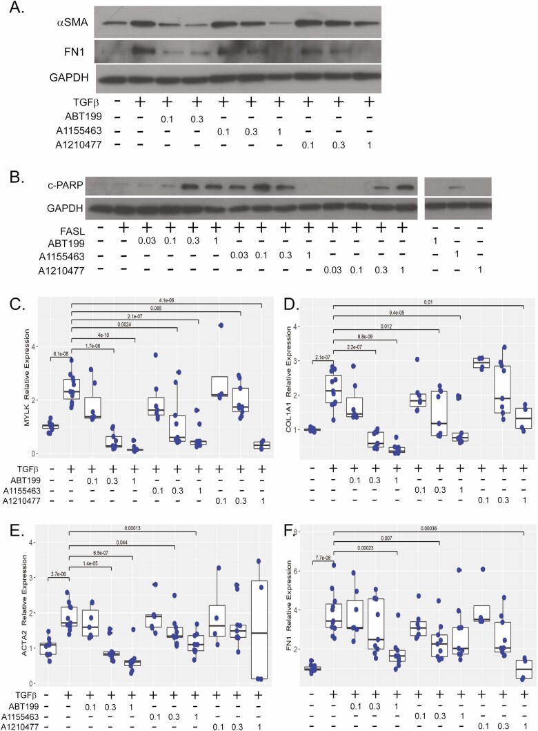
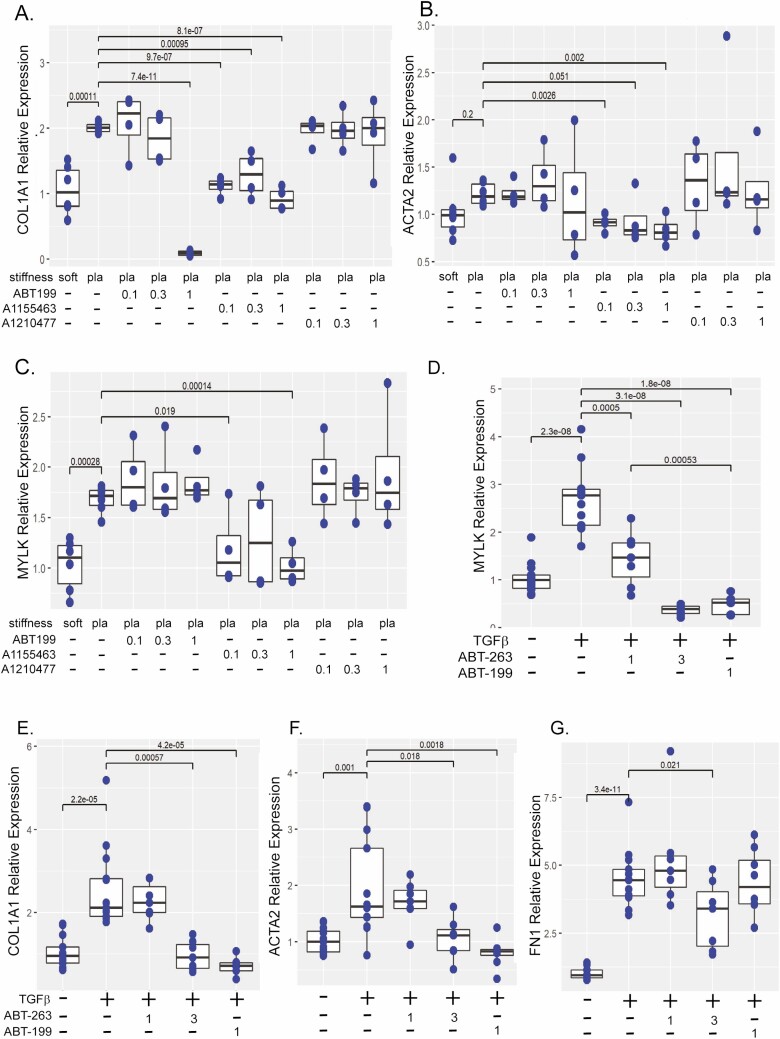
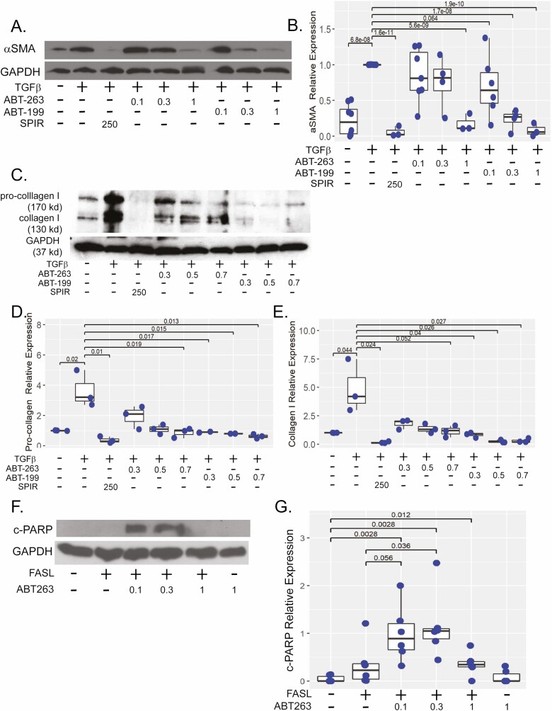
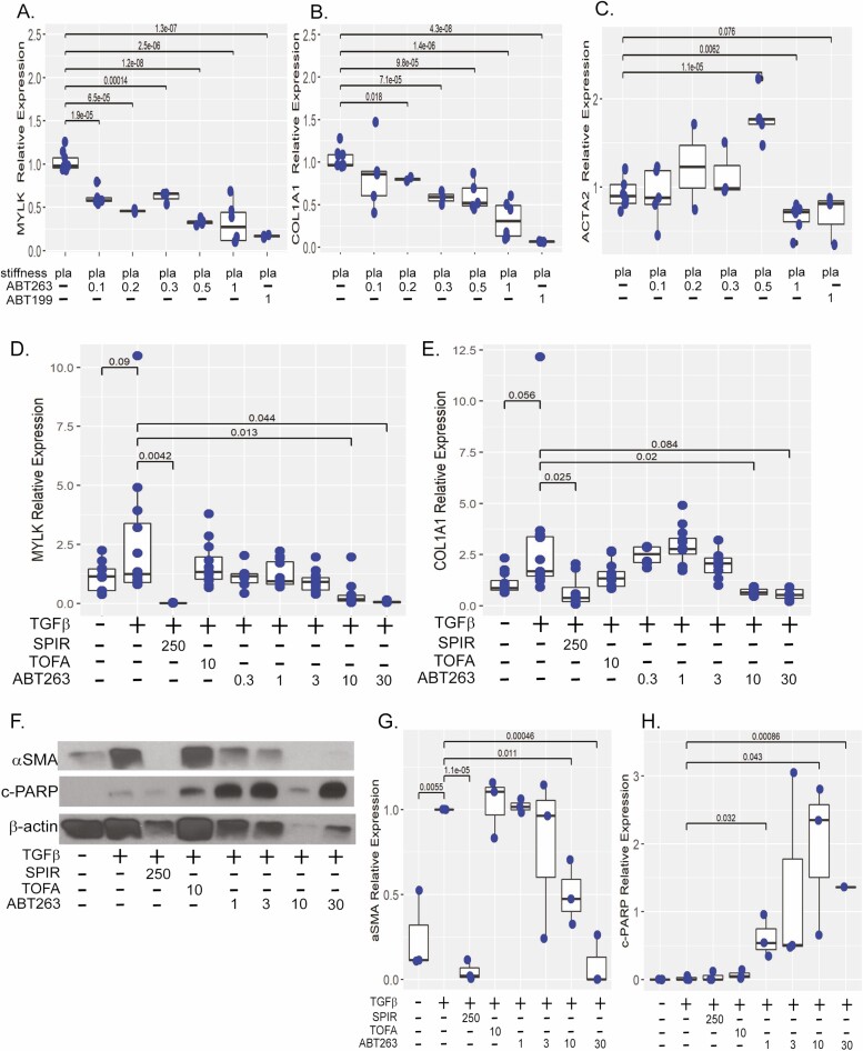
Background: Intestinal fibrosis and subsequent intestinal obstruction are common complications of Crohn's disease (CD). Current therapeutics combat inflammation, but no pharmacological therapy exists for fibrostenotic disease. Pathological persistence of activated intestinal myofibroblasts is a key driver of fibrosis in CD. In other organ systems, BH-3 mimetic drugs that affect Bcl-2 apoptotic pathways induce apoptosis in activated myofibroblasts and reduce fibrogenic gene expression, thereby reducing fibrosis.

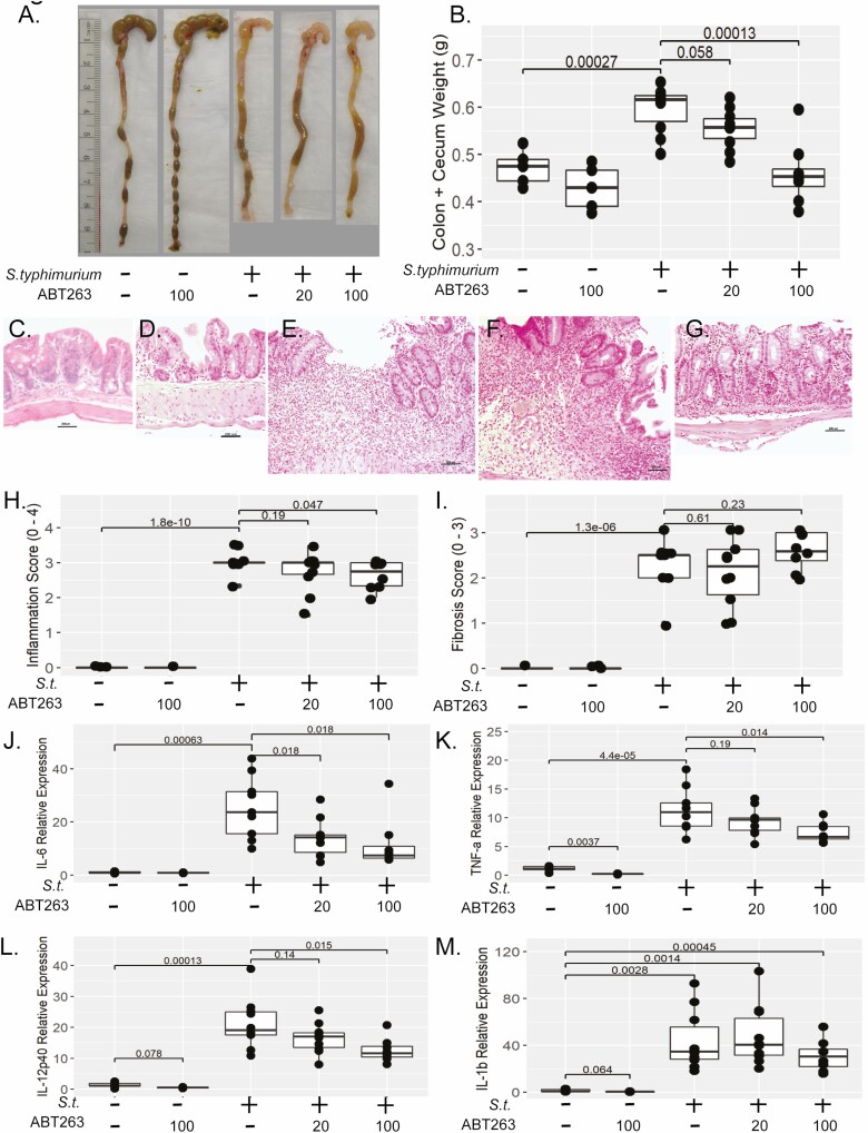
Methods: We evaluated the proapoptotic and antifibrotic efficacy of several classes of BH-3 mimetics in 2 in vitro fibrogenesis models. The candidate molecule, ABT-263, was advanced to a 3-dimensional human intestinal organoid (HIO) model. Finally, the therapeutic efficacy of ABT-263 was evaluated in the mouse Salmonella typhimurium intestinal fibrosis model.

Results: The BH-3 mimetics induced apoptosis, repressed fibrotic protein expression, and reduced fibrogenic gene expression in normal human intestinal myofibroblasts. The BH-3 mimetics that target Bcl-2 and Bcl-xl demonstrated the greatest efficacy in vitro. The ABT-199 and ABT-263 induced apoptosis and ameliorated fibrogenesis in the in vitro myofibroblast models. In the HIO model, ABT-263 inhibited fibrogenesis and induced apoptosis. In the mouse S. typhimurium model, dose-dependent reduction in macroscopic pathology, histological inflammation, inflammatory and fibrotic gene expression, and extracellular matrix protein expression indicated ABT-263 may reduce intestinal fibrosis.

Conclusions: In vitro, the antifibrotic efficacy of BH-3 mimetics identifies the Bcl-2 pathway as a druggable target and BH-3 mimetics as putative therapeutics. Reduction of inflammation and fibrosis in the mouse intestinal fibrosis model by ABT-263 indicates BH-3 mimetics as potential, novel antifibrotic therapeutics for Crohn's disease.

Keywords: ABT-263; BH-3 mimetic; Bcl-2; Crohn’s disease; fibrosis; inflammatory bowel disease; myofibroblast; navitoclax.

Plain language summary

Intestinal fibrosis is a common complication of Crohn’s disease, yet no effective therapies exist to treat fibrostenotic disease. We report ABT-263 (navitoclax) reduces intestinal fibrosis in in vitro models and reduces inflammation and fibrosis in a mouse IBD model.


Figures

Figure 1.
Figure 1.
BH-3 mimetics reduce fibrotic protein expression, sensitize intestinal myofibroblasts to apoptosis, and inhibit fibrotic gene expression. A, Representative western illustrating αSMA and fibronectin (FN1) protein expression in CCD-18co cells stimulated with 0.05 ng/mL TGFβ and cotreated with 0.1 to 0.3 μM of ABT-199 or 0.1 to 1 uM of A1155463 or A1210477 for 48 hours. GAPDH protein expression was used a control for protein loading. B, Representative cleaved PARP (c-PARP) western of CCD-18co cells treated with anti-FAS activating antibody (FASL,100 ng/mL) and 0.03 to 1 μM of each BH-3 mimetic for 5 hourss compared with non-FASL sensitized cells (untreated or 1 μM of each BH-3 mimetic). The GAPDH expression was used a control for protein loading. All lanes are from the same western and exposure. Intervening lanes from a compound outside the scope of this study have been removed for clarity. Gene expression of MYLK (C), COL1A1 (D), ACTA2 (E), and FN1 (F) in CCD-18co cells treated with 0.05 ng/mL TGFβ and cotreated with 0.1 to 3 μM of ABT-199, A1155463, or A1210477 for 24 hours compared with untreated CCD-18co cells. Fibrogenic gene expression was normalized to GAPDH expression. Results are from 4 independent experiments with 2 to 3 technical replicates per experiment. Individual replicates are represented by points within boxplots denoting the median and interquartile range. Significant (P < .05) statistical comparisons are enumerated within brackets.
Figure 2.
Figure 2.
BH-3 mimetics reduce fibrogenic gene expression in the in vitro matrix stiffness and TGF-β models. Gene expression of COL1A1 (A), ACTA2 (B), and MYLK (C) in CCD-18co cells cultured on physiologically soft (4.3 kPa) or pathologically stiff (plastic) matrices and treated with 0.1 to 1 μM of ABT-199, A1155463, or A1210477 for 48 hours compared with untreated CCD-18co cells cultured on soft or stiff matrices. Results are from 2 independent experiments with 3 biological replicates per experiment. (D–G) Comparison of ABT-263 and ABT-199 effects on gene expression of MYLK (D), COL1A1 (E), ACTA2 (F), and FN1 (G) in CCD-18co cells treated with 0.05 ng/mL TGFβ and cotreated with 1 μM or 3 μM of ABT-263 or 1 μM of ABT-199 for 24 hours compared with untreated CCD-18co cells. Results are from 4 independent experiments with 2 to 3 biological replicates per experiment. In both in vitro models, fibrogenic gene expression was normalized to GAPDH expression. Individual replicates are represented by points within boxplots denoting the median and interquartile range. Significant (P < .05) statistical comparisons are enumerated within brackets.
Figure 3.
Figure 3.
ABT-263 reduces αSMA protein expression and sensitizes intestinal myofibroblasts to apoptosis. A, Representative western illustrating αSMA protein expression in CCD-18co cells stimulated with 0.05 ng/mL TGFβ and cotreated with 0.1 to 1 μM of ABT-263 or ABT-199 48 hours compared with untreated cells or cells treated with 250 μM spironolactone, a previously described antifibrotic compound. B, Quantification of αSMA protein as normalized to GAPDH expression. Results are from 7 independent experiments. C, Representative western illustrating procollagen 1 and collagen 1 protein expression in CCD-18co cells stimulated with 0.05 ng/mL TGFβ and cotreated with 0.3 to 0.7 μM of ABT-263 or ABT-199 for 48 hours compared with untreated cells or cells treated with 250 μM of spironolactone. D, E, Quantification of procollagen and collagen 1 (normalized to GAPDH) expression. Results are from 3 independent experiments. F, Representative c-PARP western of CCD-18co cells treated with anti-FAS activating antibody (FASL, 100 ng/mL) and 0.1 to 1 μM of ABT-263 or ABT-199 for 5 hourss compared with non-FASL sensitized cells. G, Quantification of c-PARP as normalized to GAPDH protein expression. Results are from 3 independent experiments. In all experiments, GAPDH protein expression was used a control for protein loading. Individual replicates are represented by points within boxplots denoting the median and interquartile range. Significant (P < .05) statistical comparisons are enumerated within brackets.
Figure 4.
Figure 4.
ABT-263 represses matrix stiffness-induced fibrosis in myofibroblasts and HIOs. Gene expression of MYLK (A), COL1A1 (B), and ACTA (C) in CCD-18co cells cultured on pathologically stiff (plastic) matrix and treated with 0.1 to 1 μM of ABT-263 for 48 hours compared with 1 μM of ABT-199 treated or untreated CCD-18co cells. Fibrogenic gene expression was normalized to GAPDH expression. Results are from 3 independent experiments with 3 biological replicates per experiment. D–H, Human intestinal organoids HIOs were stimulated with 2 ng/mL TGFβ and cotreated with 0.3 to 30 μM of ABT-263 for 96 hours. Antifibrogenic response was compared with TGF-β-stimulated HIOs cotreated with 250 μM of spironolactone (antifibrotic, positive drug control) or 10 μM of tofacitinib (anti-inflammatory, negative drug control) and untreated HIOs (no drug, negative control). Gene expression of MYLK (D) and COL1A1 (E), normalized to GAPDH expression. Results are from 4 independent experiments with 3 biological replicates per experiment. F, Representative western demonstrating αSMA and c-PARP protein expression in HIOs stimulated with 2 ng/mL TGFβ and cotreated with 1 to 30 μM of ABT-263 for 96 hours. Quantification of αSMA (G) and c-PARP (H) as normalized to β-actin expression. Results are from 3 independent experiments. Individual replicates are represented by points within boxplots denoting the median and interquartile range. Significant (P < .05) statistical comparisons are enumerated within brackets.
Figure 5.
Figure 5.
ABT-263 reduces bowel inflammation in vivo. A, Representative macroscopic pathology of mouse cecum and distal colon weight in mice infected with S. typhimurium and treated daily with 20 or 100 mg/kg/day ABT-263 compared with S. typhimurium-infected and uninfected controls (no drug or 100 mg/kg/day ABT-263). B, Dose-dependent reduction in cecal and colon tissue weight. C–G, Photomicrographs (100x magnification) of H&E stained sections of mouse cecum of (C) uninfected, no drug. No lesions are present. D, Uninfected, drug control (100 mg/kg/day ABT-263). No lesions are present. E, S. typhimurium infected. There is a large chronic ulcer containing a dense accumulation of neutrophils extending through the lamina propria and submucosa. Adjacent cecal glands are hyperplastic. F, S. typhimurium infected treated with 20 mg/kg/day of ABT-263, similar to (E); there is epithelial erosion and hyperplasia accompanied by marked neutrophilic infiltration that extends into the submucosa. G, S. typhimurium infected treated with 100 mg/kg/day ABT-263. Inflammation and hyperplasia are moderate, and inflammation is limited to the lamina propria The scale bar denotes 200 μm. H, Summary of histological inflammation of H&E stained sections (blinded scoring, 0–4 point scale). I, Summary of histological fibrosis of Masson’s trichrome stained sections (blinded scoring, 3-point scale). Inflammatory gene expression of IL-6 (J), TNFα (K), IL-12p40 (L), and IL-1β (M) in cecal tissue from mice infected with S. typhimurium and treated daily with 20 or 100 mg/kg/day of ABT-263 compared with S. typhimurium-infected and uninfected controls (no drug or 100 mg/kg/day of ABT-263). Inflammatory gene expression was normalized to GAPDH gene expression. Individual animals are represented by points within boxplots denoting the median and interquartile range. Statistical comparisons are enumerated within brackets with P values denoted.
Figure 6.
Figure 6.
ABT-263 Reduces fibrotic gene and protein expression in vivo. Expression of fibrogenic genes CTGF (A), TGFβ (B), and IGF-1 (C) in mice infected with S. typhimurium and treated daily with 20 or 100 mg/kg/day of ABT-263 compared with S. typhimurium-infected and uninfected controls (no drug or 100 mg/kg/day of ABT-263) as normalized to GAPDH gene expression. Representative westerns blots of collagen 1 (D) and αSMA (E). GAPDH expression was used a control for protein loading. E, Quantification of collagen 1 (F) and αSMA (G) normalized to GAPDH expression. Individual animals are represented by points within boxplots denoting the median and interquartile range. Statistical comparisons are enumerated within brackets with P values denoted.

References

    1. Loftus CG, Loftus EV Jr, Harmsen WS, et al. . Update on the incidence and prevalence of Crohn’s disease and ulcerative colitis in Olmsted County, Minnesota, 1940-2000. Inflamm Bowel Dis. 2007;13:254–261. - PubMed
    1. Rieder F, Fiocchi C, Rogler G. Mechanisms, management, and treatment of fibrosis in patients with inflammatory bowel diseases. Gastroenterology. 2017;152:340–350.e6. - PMC - PubMed
    1. Li C, Kuemmerle JF. The fate of myofibroblasts during the development of fibrosis in Crohn’s disease. J Dig Dis. 2020;21:326–331. - PubMed
    1. Hinz B. The role of myofibroblasts in wound healing. Curr Res Transl Med. 2016;64:171–177. - PubMed
    1. Horowitz JC, Thannickal VJ. Mechanisms for the resolution of organ fibrosis. Physiology (Bethesda). 2019;34:43–55. - PMC - PubMed

Publication types