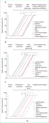
Biomarkers of conversion to α-synucleinopathy in isolated rapid-eye-movement sleep behaviour disorder
- PMID: 34302789
- PMCID: PMC8600613
- DOI: 10.1016/S1474-4422(21)00176-9
Biomarkers of conversion to α-synucleinopathy in isolated rapid-eye-movement sleep behaviour disorder
Abstract
Patients with isolated rapid-eye-movement sleep behaviour disorder (RBD) are commonly regarded as being in the early stages of a progressive neurodegenerative disease involving α-synuclein pathology, such as Parkinson's disease, dementia with Lewy bodies, or multiple system atrophy. Abnormal α-synuclein deposition occurs early in the neurodegenerative process across the central and peripheral nervous systems and might precede the appearance of motor symptoms and cognitive decline by several decades. These findings provide the rationale to develop reliable biomarkers that can better predict conversion to clinically manifest α-synucleinopathies. In addition, biomarkers of disease progression will be essential to monitor treatment response once disease-modifying therapies become available, and biomarkers of disease subtype will be essential to enable prediction of which subtype of α-synucleinopathy patients with isolated RBD might develop.
Copyright © 2021 Elsevier Ltd. All rights reserved.
Conflict of interest statement
Declaration of interests CHA reports personal fees from Acadia, Acorda, Amneal, Axovant, Jazz, Lundbeck, Neurocrine, Scion, and Sunovion, outside the submitted work. DA reports consulting fees from Fidia and Bioprojet. BFB reports personal fees from the Scientific Advisory Board-Tau Consortium and grants from Biogen, NIH, the Mangurian Foundation, Alector, the Little Family Foundation, the Turner Family, and EIP Pharma, outside the submitted work. PD reports grants from the Czech Ministry of Health, the Czech Science Foundation, the EU's Horizon 2020 research and innovation programme, and personal fees from BenevolentAI Bio, Retrophin, and Alexion Pharmaceuticals, outside the submitted work. RF reports grants from the Italian Ministry of Health, during the conduct of the study. J-FG reports grants from the Canadian Institutes of Health Research and the Canada Research Chair, during the conduct of the study. ZG-O reports grants from the Michael J Fox Foundation, Parkinson Canada, the Canadian Consortium on Neurodegeneration in Aging, the Canada First Research Excellence Fund, and personal fees from Lysosomal Therapeutics, Idorsia, Prevail Therapeutics, Deerfield, Inception Sciences (Ventus), Denali, Ono Therapeutics, and Neuron23, outside the submitted work. BH reports personal fees from Axovant, Roche, Takeda, Jazz, BenevolentAI Bio, ONO, AOP Orphan, Inspire, and Novartis, and travel support from Habel Medizintechnik Vienna, during the conduct of the study, and has a patent (“Detection of Leg Movements II” Österreichische Patentanmeldung A50649/2019) pending. MTH reports grants from Parkinson's UK, Oxford Biomedical Research Centre, MJFF, H2020 EU, GE Healthcare, and the PSP association, and personal fees from Biogen, Roche, Curasen Pharmaceuticals Consultancy Honoraria, outside the submitted work. AJ reports a grant from ParkinsonFonds Deutschland, outside the submitted work. CL reports grants from the Oxford NIHR Biomedical Research Centre, outside the submitted work. GP reports personal fees from UCB, Bioprojet, Jazz Pharma, and Idorsia, outside the submitted work. FP reports personal fees from Vanda Pharmaceuticals, Sanofi, Zambon, Fidia, Bial, Eisai Japan, and Italfarmaco, outside the submitted work. MR reports grants from NIHR, during the conduct of the study, and grants from Britannia Pharmaceuticals and Eli Lilly, and non-financial support from IXICO, outside the submitted work. WHO reports grants from ParkinsonFonds Deutschland, the Michael J Fox Foundation, and Deutsche Forschungsgemeinschaft (DFG), during the conduct of the study, and personal fees from Adamas, MODAG, Roche, and UCB, outside the submitted work, and is Hertie-Senior-Research Professor supported by the Charitable Hertie-Foundation. All other authors declare no competing interests.
Figures


Comment in
-
Identifying the best biomarkers for α-synucleinopathies.Lancet Neurol. 2021 Aug;20(8):593-594. doi: 10.1016/S1474-4422(21)00201-5. Lancet Neurol. 2021. PMID: 34302778 No abstract available.
References
-
- Ferri R, Aricò D, Cosentino FII, Lanuzza B, Chiaro G, Manconi M. REM sleep without atonia with REM sleep-related motor events: broadening the spectrum of REM sleep behavior disorder. Sleep 2018; 41: zsy187. - PubMed
-
- Högl B, Stefani A, Videnovic A. Idiopathic REM sleep behaviour disorder and neurodegeneration—an update. Nat Rev Neurol 2018; 14: 40–55. - PubMed
-
- Nepozitek J, Dostalova S, Dusek P, et al. Simultaneous tonic and phasic REM sleep without atonia best predicts early phenoconversion to neurodegenerative disease in idiopathic REM sleep behavior disorder. Sleep 2019; 42: zsz132. - PubMed
Publication types
MeSH terms
Substances
Grants and funding
LinkOut - more resources
Full Text Sources
Medical

