Review
. 2021 Oct;18(5):693-712.
doi: 10.1007/s13770-021-00373-w. Epub 2021 Jul 25.

Mucosal Vaccine Delivery Using Mucoadhesive Polymer Particulate Systems

Affiliations
Review

Mucosal Vaccine Delivery Using Mucoadhesive Polymer Particulate Systems

Chong-Su Cho et al. Tissue Eng Regen Med. 2021 Oct.

Abstract

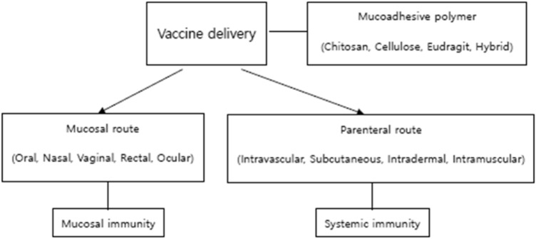
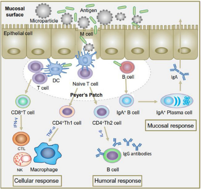
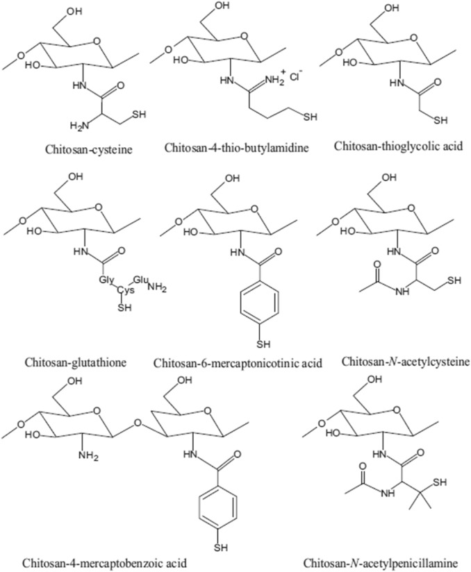
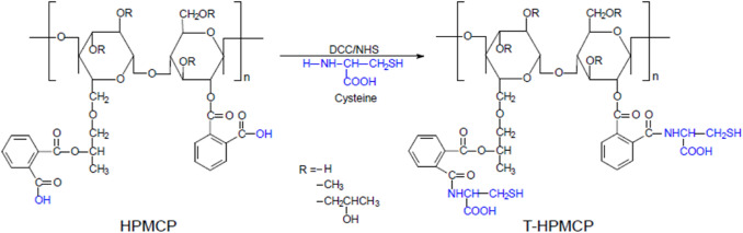
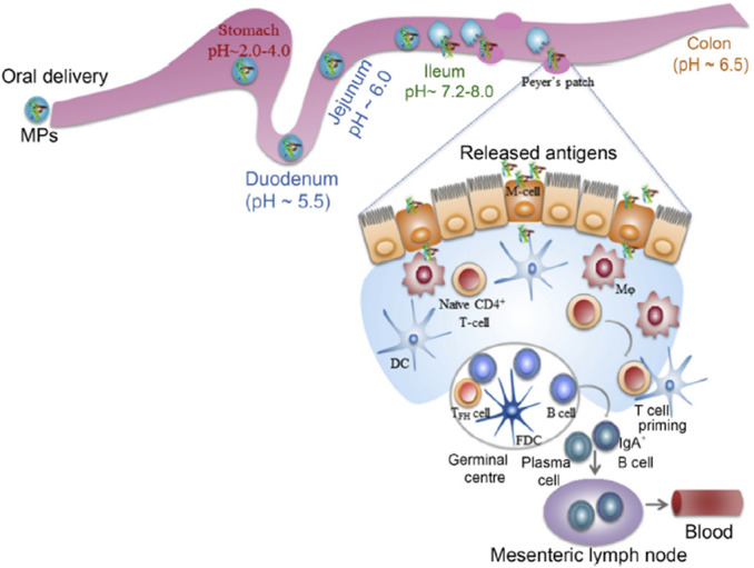
Vaccination has been recently attracted as one of the most successful medical treatments of the prevalence of many infectious diseases. Mucosal vaccination has been interested in many researchers because mucosal immune responses play part in the first line of defense against pathogens. However, mucosal vaccination should find out an efficient antigen delivery system because the antigen should be protected from degradation and clearance, it should be targeted to mucosal sites, and it should stimulate mucosal and systemic immunity. Accordingly, mucoadhesive polymeric particles among the polymeric particles have gained much attention because they can protect the antigen from degradation, prolong the residence time of the antigen at the target site, and control the release of the loaded vaccine, and results in induction of mucosal and systemic immune responses. In this review, we discuss advances in the development of several kinds of mucoadhesive polymeric particles for mucosal vaccine delivery.

Keywords: Antigen delivery; Mucoadhesive polymers; Mucosal vaccination; Polymeric particles.


Conflict of interest statement

The authors have no financial conflicts of interest.

Figures

Fig. 1
Fig. 1
Schematic illustration of the contents
Fig. 2
Fig. 2
Schematic diagram of various immune responses induced by particulate vaccine system. Upon encounter with an antigen, B cells convert themselves to antibody secreting plasma cells that produce antibodies for excreting the pathogens to mucosal surfaces (mucosal response) whereas dendritic cells (DCs) present the antigen via major histocompatibility complex (MHC) class I and class II molecules to CD8 + and CD4 + T-cells. Activation pathway of CD8 + T cells and CD4 + Th1 cells produces cytotoxicT lymphocytes (CTL) and activated macrophages that kill intracellular pathogens or infected cells (cellular response) while activation pathway of CD4 + Th2 cells produce sactivated B lymphocytes that secrete antibodies for neutralization of extracellular pathogens (humoral response). Adapted from Singh et al., Chitosan-based particulate systems for the delivery of mucosal vaccines against infectious diseases. International Journal of Biological Macromolecules 2018, 110, 54–64, with permission of Elsevier [11]
Fig. 3
Fig. 3
Chemical structures of the presented chemical-modified chitosan variants. Adapted from Islam et al., Mucoadhesive Chitosan Derivatives as Novel Drug Carriers, Current Pharmaceutical Design, 2015, 21, 4285–4309 with permission of Bentham Science [30]
Fig. 4
Fig. 4
The reaction scheme for the synthesis of T-HPMCP. Adapted from Singh et al., Attuning hydroxypropyl methylcellulose phthalate to oral delivery vehicle for effective and selective delivery of protein vaccine in ileum. Biomaterials 2015, 59, 144 ~ 159 with permission of Elsevier [52]
Fig. 5
Fig. 5
Design for oral delivery of vaccines targeted to M cells in ileum. Intraluminal pH and GI transit time are indicated (distance not to scale). Microparticles (MPs) are expected to begin to dissolve in the ileum for uptake of released antigens through M cells. Adapted Grabovac et al., Comparison of the mucoadhesive properties of various polymers. Adv Drug Deliv Rev 2005, 57, 1713 ~ 1723 with permission of Elsevier [53]
Fig. 6
Fig. 6
Analysis of morphology and size of MPs. Morphology of the MPs was analyzed by SEM (scale bar: 2 mm). FITC-labeled antigen/MPs were observed by CLSM. A M-BmpB/THPMCP MPs and FITC-M-BmpB/T-HPMCP MPs (inset); B M-BmpB/HPMCP MPs and FITC-M-BmpB/HPMCP MPs (inset). The particle-size distributions were detected by DLS. C MBmpB/T-HPMCP MPs; D M-BmpB/HPMCP MPs. Adapted from Singh et al., Attuning hydroxypropyl methylcellulose phthalate to oral delivery vehicle for effective and selective delivery of protein vaccine in ileum. Biomaterials 2015, 59, 144–159 with permission of Elsevier [53]
Fig. 7
Fig. 7
Localization of FITC-labeled M-BmpB in Peyer's patch of mouse small intestine. A FITC-labeled M-BmpB/T-HPMCP or M-BmpB/HPMCP MPs were orally administered into the mice and their localization was monitored under fluorescence-microscopy. The green fluorescent signals of FITC-labeled M-BmpB, when delivered by T-HPMCP MPs, were higher in Peyer's patch underneath the FAE region. B Uptake of FITC-M-BmpB was quantitated by image J analysis and normalized to a value of 1.0 for M-BmpB control. Scale bar: 200 mm. (For interpretation of the references to color in this figure legend, the reader is referred to the web version of this article.) Adapted from Singh et al., Attuning hydroxypropyl methylcellulose phthalate to oral delivery vehicle for effective and selective delivery of protein vaccine in ileum. Biomaterials 2015, 59, 144 ~ 159 with permission of Elsevier [52]
Fig. 8
Fig. 8
A Confocal microscopic images of RAW264.7 cells after 2 h culture with OVA-loaded THMand OVA-loaded Man-THMat 4 °C and 37 °C. B Measurement of microsphere uptake by RAW264.7 using FACS. Uptake of OVA-FITC-loaded THM and OVA-FITC loaded Man-THM by RAW 264.7 in 1 h and 2 h with and without MR inhibition at 37 °C (n = 3, error bar represents standard deviation; *p < 0.05, **p < 0.01, ***p < 0.005, one-way ANOVA). Adapted from Li et al., Nasal immunization with mannan-decorated mucoadhesive HPMCP microspheres containing ApxIIA toxin induces protective immunity against challenge infection with Actinobacillus pleuropneumoiae in mice. Journal of Controlled Release 2016, 233, 114–125 with permission of Elsevier [54]
Fig. 9
Fig. 9
AC ApxIIA-specific IgA performance in themucosal sites at 4 weeks post-immunization. ApxIIA-specific brochealveolar lavage (A), nasal wash (B), vaginal wash (C), IgA levels inmice immunizedwith the indicated formulationswere analyzed by ELISA and then calculated by optical density (450 nm) (n = 5, error bars represent standard deviations; *p < 0.05, **p < 0.01, ***p < 0.005, one-way ANOVA). Adapted from Li et al., Nasal immunization with mannan-decorated mucoadhesive HPMCP microspheres containing ApxIIA toxin induces protective immunity against challenge infection with Actinobacillus pleuropneumoiae in mice. Journal of Controlled Release 2016, 233, 114–125 with permission of Elsevier [54]
Fig. 10
Fig. 10
Induction of protective immunity after intranasal challenge with A. pleuropneumoniae. A 14 days after the last immunization, 5mice per group were challenged intranasally with a minimal lethal dose (5 × 107 CFU) of A. pleuropneumoniae,with the survival rate (%)monitored for an additional 4 days. B The number of residual bacteria was counted per 100mgfresh lung tissue weight from each mice per group. (p < 0.05, **p < 0.01, ***p < 0.005, one-way ANOVA) C The lungs were characterized before becoming homogenates and after bacteria challenge. Adapted from Li et al., Nasal immunization with mannan-decorated mucoadhesive HPMCP microspheres containing ApxIIA toxin induces protective immunity against challenge infection with Actinobacillus pleuropneumoiae in mice. Journal of Controlled Release 2016, 233, 114–125 with permission of Elsevier [54]
Fig. 11
Fig. 11
Amount of FDA remained on excised porcine small intestinal mucosa. Adapted from Quan et al., pH-sensitive and mucoadhesive thiolated Eudragit-coated chitosan microspheres. International Journal of Pharmaceutics 2008, 359,205–210 with permission of Elsevier [65]

References

    1. Corthésy B, Bioley G. Lipid-based particles: versatile delivery systems for mucosal vaccination against infection. Front Immunol. 2018;9:431. doi: 10.3389/fimmu.2018.00431. - DOI - PMC - PubMed
    1. Yoon SY, Kang SK, Lee HB, Oh SH, Kim WS, Li HS, et al. Enhanced efficacy of immunization with a foot-and-mouth disease multi-epitope subunit vaccine using mannan-decorated inulin microparticles. Tissue Eng Regen Med. 2020;17:33–44. doi: 10.1007/s13770-019-00228-5. - DOI - PMC - PubMed
    1. Lim W, Kim HS. Exosomes as therapeutic vehicles for cancer. Tissue Eng Regen Med. 2019;16:213–223. doi: 10.1007/s13770-019-00190-2. - DOI - PMC - PubMed
    1. Pulendran B, Ahmed R. Immunological mechanisms of vaccination. Nat Immunol. 2011;12:509–517. doi: 10.1038/ni.2039. - DOI - PMC - PubMed
    1. Longet S, Lundahl MLE, Lavelle EC. Targeted strategies for mucosal vaccination. Bioconjug Chem. 2018;29:613–623. doi: 10.1021/acs.bioconjchem.7b00738. - DOI - PubMed

Publication types

LinkOut - more resources