. 2021 Sep;22(3):914.
doi: 10.3892/etm.2021.10346. Epub 2021 Jun 29.

ZFAS1 knockdown inhibits fibroblast-like synoviocyte proliferation, migration, invasion and inflammation, and promotes apoptosis via miR-3926/FSTL1 in rheumatoid arthritis

Affiliations

ZFAS1 knockdown inhibits fibroblast-like synoviocyte proliferation, migration, invasion and inflammation, and promotes apoptosis via miR-3926/FSTL1 in rheumatoid arthritis

Qiang Wang et al. Exp Ther Med. 2021 Sep.

Abstract

Rheumatoid arthritis (RA) is a chronic systemic autoimmune disease characterized by joint disorders. Long non-coding RNA zinc finger antisense 1 (ZFAS1) is aberrantly expressed in numerous human diseases, including RA. The present study aimed to investigate the functions and underlying mechanisms of ZFAS1 in RA. Reverse transcription-quantitative PCR was performed to determine the expression levels of ZFAS1, microRNA (miR)-3926 and follistatin-like protein 1 (FSTL1). MTT assay, flow cytometric analysis and Transwell assay were performed to examine the proliferation, apoptosis, migration and invasion of fibroblast-like synoviocytes (FLSs), respectively. Western blotting was employed to measure the protein expression levels of cleaved caspase-3, interleukin (IL)-6, IL-1β, tumor necrosis factor-α and FSTL1. Dual-luciferase reporter assay was performed to verify the interaction between miR-3926 and ZFAS1 or FSTL1. The results demonstrated that ZFAS1 and FSTL1 were upregulated, and miR-3926 was downregulated in RA synovial tissues and RA-FLSs. ZFAS1 knockdown suppressed cell proliferation, migration, invasion and inflammatory cytokine production, and induced apoptosis in RA-FLSs. ZFAS1 acted as a sponge for miR-3926, and ZFAS1 overexpression abolished the impact of miR-3926 on the development of RA-FLSs. FSTL1 was a direct target of miR-3926, and the effect of FSTL1 knockdown on the progression of RA-FLSs was rescued by miR-3926 inhibition. Furthermore, ZFAS1 regulated FSTL1 expression levels via sponging miR-3926 in RA-FLSs. In conclusion, ZFAS1 knockdown inhibited RA-FLS proliferation, migration, invasion and inflammatory cytokine production, and induced apoptosis in RA via the miR-3926/FSTL1 axis.

Keywords: fibroblast-like synoviocytes; follistatin-like protein 1; microRNA-3926; rheumatoid arthritis; zinc finger antisense 1.


Conflict of interest statement

The authors declare that they have no competing interests.

Figures

Figure 1
Figure 1
ZFAS1 expression levels are significantly elevated in RA synovial tissues and RA-FLSs. Expression levels of ZFAS1 in (A) RA synovial tissue and non-arthritic control tissue, and (B) RA-FLSs and N-FLSs were analyzed via reverse transcription-quantitative PCR. *P<0.05. ZFAS1, zinc finger antisense 1; RA, rheumatoid arthritis; FLSs, fibroblast-like synoviocytes; N, normal.
Figure 2
Figure 2
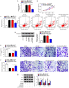
ZFAS1 downregulation suppresses cell proliferation, migration, invasion and inflammatory cytokine expression, and facilitates cell apoptosis in RA-FLSs. RA-FLSs were transfected with si-NC or si-ZFAS1. (A) Expression levels of ZFAS1 were detected via reverse transcription-quantitative PCR. (B) Proliferation of RA-FLSs was evaluated via MTT assay. (C) Apoptosis of RA-FLSs was assessed via flow cytometric analysis. (D) Protein expression levels of C-caspase-3 were measured via western blotting. (E) Migration and (F) invasion of RA-FLSs were examined via Transwell assay (magnification, x100). (G) Expression levels of IL-6, IL-1β and TNF-α were measured via western blotting. *P<0.05 vs. si-NC. ZFAS1, zinc finger antisense 1; RA-FLSs, rheumatoid arthritis-fibroblast-like synoviocytes; si, small interfering RNA; NC, negative control; C-caspase-3, cleaved-caspase-3; IL, interleukin; TNF-α, tumor necrosis factor-α; PI, propidium iodide.
Figure 3
Figure 3
ZFAS1 directly binds miR-3926 and negatively regulates miR-3926 expression levels in RA-FLSs. (A) Predicted binding sites between ZFAS1 and miR-3926. (B) Luciferase activity in RA-FLSs transfected with ZFAS1 WT or ZFAS1 MUT together with miR-3926 or miR-NC was analyzed via dual-luciferase reporter assay. Expression levels of miR-3926 in (C) RA synovial and non-arthritic control tissue, and (D) RA-FLSs and N-FLSs were assessed via RT-qPCR. (E) Correlation between expression levels of ZFAS1 and miR-3926 in RA-FLSs was analyzed via Pearson's correlation analysis. (F) Expression levels of ZFAS1 in RA-FLSs transfected with pcDNA-ZFAS1 or pcDNA-NC was examined via RT-qPCR analysis. (G) RA-FLSs were transfected with si-NC, si-ZFAS1, pcDNA-NC or pcDNA-ZFAS1, then the expression levels of miR-3926 were determined via RT-qPCR. (H) Expression levels of miR-3926 in RA-FLSs transfected with anti-miR-NC or anti-miR-3926 were determined via RT-qPCR. *P<0.05 vs. miR-NC. ZFAS1, zinc finger antisense 1; miR, microRNA; RA-FLSs, rheumatoid arthritis-fibroblast-like synoviocytes; WT, wild type; MUT, mutant; NC, negative control; N, normal; RT-qPCR, reverse transcription-quantitative PCR; si, small interfering RNA.
Figure 4
Figure 4
ZFAS1 alters cell proliferation, apoptosis, migration, invasion and inflammatory cytokine expression via binding to miR-3926 in RA-FLSs. RA-FLSs were transfected with miR-NC, miR-3926, miR-3926 + pcDNA-NC or miR-3926 + pcDNA-ZFAS1. (A) Expression levels of miR-3926 in RA-FLSs were measured via reverse transcription-quantitative PCR. (B) RA-FLS proliferation was determined via MTT assay. (C) RA-FLS apoptosis was analyzed via flow cytometric analysis. (D) Expression levels of C-caspase-3 in RA-FLSs were determined via western blotting. (E) Migration and (F) invasion of RA-FLS were assessed via Transwell assay (magnification, x100). (G) Expression levels of IL-6, IL-1β and TNF-α were determined via western blotting assay. *P<0.05. ZFAS1, zinc finger antisense 1; miR, microRNA; RA-FLSs, rheumatoid arthritis-fibroblast-like synoviocytes; NC, negative control; C-caspase-3, cleaved-caspase-3; IL, interleukin; TNF-α, tumor necrosis factor-α.
Figure 5
Figure 5
miR-3926 directly targets FSTL1 and negatively regulates its expression in RA-FLSs. (A) Potential binding sites between miR-3926 and FSTL1 were predicted via TargetScan. (B) Dual-luciferase reporter assay was performed to determine luciferase activity in RA-FLSs co-transfected with FSTL1 3'UTR WT or FSTL1 3'UTR MUT and miR-3926 or miR-NC. (C) mRNA and (D) protein expression levels of FSTL1 in RA synovial and non-arthritic control tissue were analyzed via RT-qPCR and western blotting, respectively. (E) mRNA and (F) protein expression levels of FSTL1 in RA-FLSs and N-FLSs were assessed via RT-qPCR and western blotting, respectively. (G) Pearson's correlation analysis was utilized to analyze the correlation between miR-3926 and FSTL1 in RA synovial tissue. RA-FLSs were transfected with miR-NC, miR-3926, anti-miR-NC or anti-miR-3926 and the (H) mRNA and (I) protein expression levels of FSTL1 were determined via RT-qPCR and western blotting assay, respectively. *P<0.05 vs. miR-NC. miR, microRNA; FSTL1, follistatin-like protein 1; UTR, untranslated region; WT, wild type; MUT, mutant; NC, negative control; RT-qPCR, reverse transcription-quantitative PCR; RA, rheumatoid arthritis; FLSs, fibroblast-like synoviocytes; N, normal.
Figure 6
Figure 6
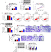
Inhibition of miR-3926 restores the inhibitory effect of FSTL1 knockdown on RA-FLSs development. RA-FLSs were transfected with si-NC, si-FSTL1, si-FSTL1 + anti-miR-NC or si-FSTL1 + anti-miR-3926. (A) mRNA and (B) protein expression levels of FSTL1 in RA-FLSs were determined via reverse transcription-quantitative PCR and western blotting, respectively. (C) Cell proliferation in RA-FLSs was assessed via MTT assay. (D) Cell apoptosis in RA-FLSs was evaluated via flow cytometric analysis. (E) Expression levels of C-caspase-3 in RA-FLSs were determined via western blotting. Cell (F) migration and (G) invasion of RA-FLSs were determined by Transwell assay (magnification, x100). (H) Expression levels of IL-6, IL-1β and TNF-α in RA-FLSs were assessed via western blotting. *P<0.05. miR, microRNA; FSTL1, follistatin-like protein 1; RA-FLSs, rheumatoid arthritis-fibroblast-like synoviocytes; si, small interfering RNA; NC, negative control; C-caspase-3, cleaved-caspase-3; IL, interleukin; TNF-α, tumor necrosis factor-α.
Figure 7
Figure 7
ZFAS1 positively modulates FSTL1 expression via miR-3926 in RA-FLSs. (A) Correlation between ZFAS1 and FSTL1 in RA synovial tissue was analyzed via Pearson's correlation analysis. (B) mRNA and (C) protein expression levels of FSTL1 in RA-FLSs transfected with si-NC, si-ZFAS1, si-ZFAS1 + anti-miR-NC or si-ZFAS1 + anti-miR-3926 were assessed via reverse transcription quantitative PCR and western blotting, respectively. *P<0.05. ZFAS1, zinc finger antisense 1; FSTL1, follistatin-like protein 1; miR, microRNA; RA-FLSs, rheumatoid arthritis-fibroblast-like synoviocytes; si, small interfering RNA; NC, negative control.

References

    1. Araki Y, Mimura T. The mechanisms underlying chronic inflammation in rheumatoid arthritis from the perspective of the epigenetic landscape. J Immunol Res. 2016;2016(6290682) doi: 10.1155/2016/6290682. - DOI - PMC - PubMed
    1. Torre LA, Bray F, Siegel RL, Ferlay J, Lortet-Tieulent J, Jemal A. Global cancer statistics, 2012. CA Cancer J Clin. 2015;65:87–108. doi: 10.3322/caac.21262. - DOI - PubMed
    1. Bottini N, Firestein GS. Duality of fibroblast-like synoviocytes in RA: Passive responders and imprinted aggressors. Nat Rev Rheumatol. 2013;9:24–33. doi: 10.1038/nrrheum.2012.190. - DOI - PMC - PubMed
    1. McInnes IB, Schett G. The pathogenesis of rheumatoid arthritis. N Engl J Med. 2011;365:2205–2219. doi: 10.1056/NEJMra1004965. - DOI - PubMed
    1. Brennan FM, McInnes IB. Evidence that cytokines play a role in rheumatoid arthritis. J Clin Invest. 2008;118:3537–3545. doi: 10.1172/JCI36389. - DOI - PMC - PubMed