. 2021 Jun 15;13(6):5985-6000.
eCollection 2021.

PLAC8 gene knockout increases the radio-sensitivity of xenograft tumors in nude mice with nasopharyngeal carcinoma by promoting apoptosis

Affiliations

PLAC8 gene knockout increases the radio-sensitivity of xenograft tumors in nude mice with nasopharyngeal carcinoma by promoting apoptosis

Li-Jun Shen et al. Am J Transl Res. .

Abstract

In vitro cell experiments showed that knocking out the placenta-specific protein 8 (PLAC8) gene significantly increased the sensitivity of tumor cells to radiation. This study used two nude mouse models of nasopharyngeal carcinoma (NPC) to investigate the radio-sensitization and molecular mechanism of PLAC8 knockout in vivo. The expression of PLAC8 in 120 NPC tissues and 30 nasopharyngitis (NPG) tissues was detected by immunohistochemistry (IHC) to analyze the relationship between PLAC8 and neck lymph node metastasis and prognosis in NPC patients. The mRNA expression level of PLAC8 in several NPC cell lines was detected by semi-quantitative RT-PCR. The PLAC8 gene was knocked out in CNE-2 cells using CRISPR/Cas9. The effect of PLAC8 gene knockout on the radiotherapy sensitivity of NPC cells was analyzed by establishing model 1 and model 2 tumor-bearing nude mouse models with two different irradiation methods. The expression of γH2AX, Bax, Bcl-2, Caspase-3 and cleaved Caspase-3 was detected by immunofluorescence (IF), IHC and western blot analysis. PLAC8 expression was significantly increased in NPC tissue samples and NPC cell lines compared with NPG tissue samples and normal cell lines (P<0.01). PLAC8 upregulation was associated with lymph node metastasis and a poor prognosis in patients with NPC (P<0.01). Both animal models showed that radiotherapy after PLAC8 knockout significantly slowed tumor growth and reduced tumor volume, with tumor inhibition rates of 100% and 66.04%, respectively. In model 2, PLAC8 knockout with radiotherapy increased the expressions of γH2AX, Bax, Caspase-3 and cleaved Caspase-3 but decreased the expression of Bcl-2 (P<0.01). In model 1, there was no tumor formation at the site where the cancer cells were injected. The expression levels of γH2AX, Bax, Caspase-3 and cleaved Caspase-3 in skin tissues taken at the injection site were lower than those in NPC tissues treated with radiotherapy, while the expression level of Bcl-2 was higher (P<0.01). PLAC8 expression is closely related to neck metastasis and the prognosis of NPC. PLAC8 gene knockout significantly increases the radio-sensitivity of NPC cells in vivo by promoting apoptosis, which is an effective strategy for the radiotherapy sensitization of NPC.

Keywords: NPC; PLAC8; apoptosis; radio-sensitivity; subcutaneously transplanted tumor model.


Conflict of interest statement

None.

Figures

Figure 1
Figure 1
Expression of the PLAC8 protein in NPC tissues and PLAC8 mRNA in NPC cell lines. (A) IHC results of tissue samples from patients with NPG and NPC. (a/b and c/d) indicate NPG negative expression and NPC strong positive expression, respectively. Image magnification in (a/c and b/d): ×100 and ×400, respectively. (B) PLAC8 expression in NPC tissues was higher than that in NPG tissues (n=150, ***P<0.001). (C) RT-PCR showed higher PLAC8 mRNA levels in human NPC cell lines (CNE-2, HK1, SUNE1, 5-8F, and 6-10B) than in NP69 cell lines. Data are shown as the mean ± standard error of the mean of three technical replicates (*P<0.05).
Figure 2
Figure 2
Expression of PLAC8 in NPC tissues and its relationship with clinical characteristics. (A) IHC results of tissue samples from NPC patients without neck lymph node metastasis. (a and b) are the IHC results of tissues without neck lymph node metastasis, while (c and d) are the IHC results of tissues with neck lymph node metastasis. Image magnification in (a/c and b/d): ×100 and ×400, respectively. (B) Quantitative analysis of PLAC8 in NPC tissues. (C) PLAC8 was upregulated in the majority of NPC patients with neck metastasis (n=120, **P<0.01). (D) Survival curves suggest that patients with NPC and low PLAC8 expression have a prolonged survival time (n=120, P<0.01).
Figure 3
Figure 3
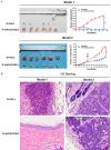
Both models confirmed that knockout of the PLAC8 gene increased the radio-sensitivity of NPC cells. After exposure to both irradiation methods, complete tumors were obtained from the IR+CNE-2 and IR+koPLAC8 CNE-2 tumor-bearing nude mouse models. A. In model 1, the tumors of nude mice in the IR+koPLAC8 group disappeared, and the tissue at the inoculation site (shown) was collected. In model 1 and model 2, there was a significant difference in tumor volume after irradiation (n=6, *P<0.05, **P<0.01). B. In model 1, the tissue at the inoculation site was confirmed as skin tissue by HE staining. Image magnification: ×200.
Figure 4
Figure 4
Expression of γH2AX, Bax, Bcl-2, Caspase-3 and cleaved Caspase-3. A. Protein staining with IF in the IR+CNE-2 and IR+koPLAC8 CNE-2 groups in model 1. B. Protein staining with IF in the IR+CNE-2 and IR+koPLAC8 CNE-2 groups in model 2. C. In model 1, compared with the fluorescence intensity in the tumor tissues of the IR+CNE-2 group, the skin tissues of the IR+koPLAC8 CNE-2 group had weaker fluorescence of γH2AX, Bax, Caspase-3 and cleaved Caspase-3, while that of Bcl-2 was stronger. D. In model 2, compared with the fluorescence intensities in the IR+CNE-2 group, those of γH2AX, Bax, Caspase-3 and cleaved Caspase-3 in the IR+koPLAC8 CNE-2 group were stronger, while that of Bcl-2 was weaker (red fluorescence indicates positive expression, while blue fluorescence indicates DAPI staining). Image magnification: ×200. *P<0.05, **P<0.01, ***P<0.001.
Figure 5
Figure 5
Staining and quantitative analysis of γH2AX, Bax, Bcl-2, Caspase-3 and cleaved Caspase-3 expression under an optical microscope. A. Protein staining in the IR+CNE-2 and IR+koPLAC8 CNE-2 groups in model 1. B. Protein staining in the IR+CNE-2 and IR+koPLAC8 CNE-2 groups in model 2. C. The expression of γH2AX, Bax, Caspase-3 and cleaved Caspase-3 was significantly lower in the IR+koPLAC8 CNE-2 group than that in the IR+CNE-2 group, while that of Bcl-2 was significantly higher. D. The expression of γH2AX, Bax, Caspase-3 and cleaved Caspase-3 was significantly higher in the IR+koPLAC8 CNE-2 group than that in the IR+CNE-2 group, while that of Bcl-2 was lower (brown-yellow particles indicate positive staining, while blue indicates nuclear staining). Image magnification: ×200. *P<0.05, **P<0.01, ***P<0.001.
Figure 6
Figure 6
Knockout of PLAC8 enhanced radio-sensitivity by activating the apoptotic pathway. A. Target protein bands obtained by western blot analysis and their statistical graph in model 1. B. Target protein bands obtained by western blot analysis and their statistical graph in model 2. Data are shown as the mean ± standard error of the mean of three technical replicates (*P<0.05, **P<0.01).

References

    1. Chen YP, Chan ATC, Le QT, Blanchard P, Sun Y, Ma J. Nasopharyngeal carcinoma. Lancet. 2019;394:64–80. - PubMed
    1. Carioli G, Negri E, Kawakita D, Garavello W, La Vecchia C, Malvezzi M. Global trends in nasopharyngeal cancer mortality since 1970 and predictions for 2020: focus on low-risk areas. Int J Cancer. 2017;140:2256–2264. - PubMed
    1. Tian Y, Yan M, Zheng J, Li R, Lin J, Xu A, Liang Y, Zheng R, Yuan Y. miR-483-5p decreases the radiosensitivity of nasopharyngeal carcinoma cells by targeting DAPK1. Lab Invest. 2019;99:602–611. - PubMed
    1. Wang C, Su Z, Hou H, Li D, Pan Z, Tian W, Mo C. Inhibition of anaphase-promoting complex by silence APC/C(Cdh1) to enhance radiosensitivity of nasopharyngeal carcinoma cells. J Cell Biochem. 2017;118:3150–3157. - PubMed
    1. Lan R, Huang F, Zhong G, Chen R, Wang Z, Chen J, Fu L, Hong J, Zhang L. Effects of CKMT1 on radiosensitivity of nasopharyngeal carcinoma cells. Int J Radiat Biol. 2019;95:597–606. - PubMed

LinkOut - more resources